COVID-19

Outside of the loss of human life due to the COVID-19 pandemic, the past two years have greatly affected hospitals, health systems and the way providers deliver care. Healthcare executives are grappling with federal monetary assistance, growing burnout rates, workforce shortages and federal oversight of vaccines and testing. This channel is also designed to update clinicians on new research and guidelines regarding COVID patient treatment strategies and risk assessments.

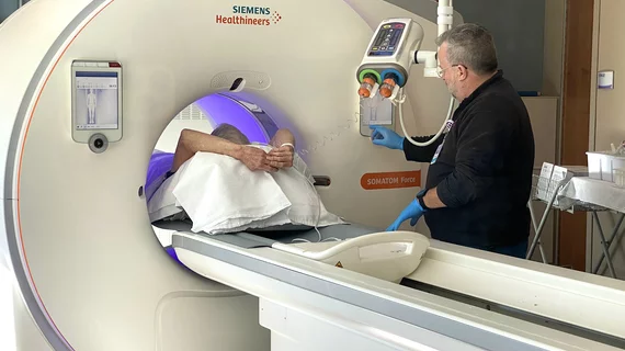
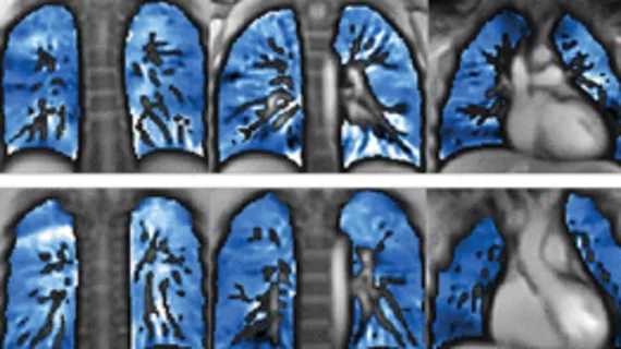
Sean Fain, PhD, vice chair of radiology and research and a professor of radiology, Division of Cardiovascular and Thoracic Imaging, University of Iowa, discusses how long-COVID lung damage can be tracked using xenon (Xe) gas magnetic resonance imaging (MRI) and quantitative computed tomography (CT). He spoke to Health Imaging at RSNA 2022.

VIDEO: Tracking long-COVID lung damage using MRI and CT

Sean Fain, PhD, vice chair of radiology and research and a professor of radiology, University of Iowa, discusses how long-COVID lung damage can be tracked using xenon (Xe) gas MRI and quantitative CT at RSNA 2022. 

Jorge Soto, MD, chair of the RSNA Annual Meeting Program Planning Committee, chief of radiology, Boston Medical Center, and professor of medicine, Boston University School of Medicine, offers an overview of the trends, hot topics, and innovative research and technology at the Radiological Society of North America (RSNA) 2022 meeting.

VIDEO: Key takeaways from RSNA 2022

Jorge Soto, MD, chair of the RSNA Annual Meeting Program Planning Committee, chief of radiology, Boston Medical Center, offers an overview of the trends, hot topics, research and technology at the Radiological Society of North America (RSNA) 2022 meeting.

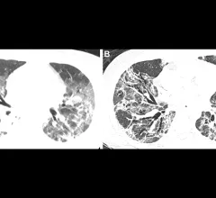
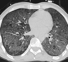
Patient with severe, lung damage from COVID. Images in a 54-year-old man with COVID-19–related acute respiratory distress syndrome and subsequent fibrosis. (A) Axial CT 2 weeks after admission shows diffuse ground-glass opacity (GGO) with reticular abnormality and traction bronchiectasis in right middle lobe, indicating organizing phase of lung injury. (B) Axial CT 6 months after admission shows decreased GGO but extensive traction bronchiectasis and architectural distortion, suggesting fibrosis. RSNA

Lung scarring occurs in up to 11% of patients recovered from COVID, regardless of infection severity

Researchers cannot yet determine whether these changes will progress or resolve in the long term.

Lung CT of a 42-year-old man with more than 7 days of symptoms. Scans show COVID pneumonia with diffuse ground-glass opacities in close vicinity of visceral pleural surfaces. In addition, a crazy paving pattern is observed. Image courtesy of RSNA. Medical images of COVID. #COVID #SARS-CoV-2

'Crash course' on COVID cases benefits residents less experienced with thoracic CT

Following the course, experts observed nearly perfect agreement between less experienced residents and chest-devoted radiologists.

MRI data reveal how pandemic-related stress physically aged the brains of teens

Researchers involved in the study suggested that, until now, this sort of growth acceleration in adolescents had only been observed in children who had experienced extreme adversity, such as violence, neglect and family dysfunction. 

Thumbnail

FDG PET/CT spots COVID-induced pancreatitis

There are limited data to determine whether pancreatitis in COVID patients is the result of a stress response triggered by severe illness or by direct damage to the β-cell structure and function.

Thumbnail

'Significant' brain abnormalities shown to persist 6 months after COVID recovery

The regions that displayed the most significant differentiations in susceptibility values are known to be associated with neurological conditions such as fatigue, insomnia, anxiety, depression, headaches and other cognitive issues—all common complaints among many COVID long haulers. 

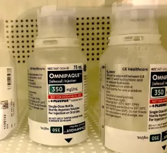
Iodinated contrast use before, during and after COVID: Trends predict significant growth in demand

A new analysis suggests that, if the use of iodinated contrast were to follow pre-pandemic trends, it could rise by as much as 37% from 2019. 

Around the web

GE HealthCare designed the new-look Revolution Vibe CT scanner to help hospitals and health systems embrace CCTA and improve overall efficiency.

Clinicians have been using HeartSee to diagnose and treat coronary artery disease since the technology first debuted back in 2018. These latest updates, set to roll out to existing users, are designed to improve diagnostic performance and user access.

The cardiac technologies clinicians use for CVD evaluations have changed significantly in recent years, according to a new analysis of CMS data. While some modalities are on the rise, others are being utilized much less than ever before.