Low-dose CT scans just as accurate as standard for diagnosing appendicitis
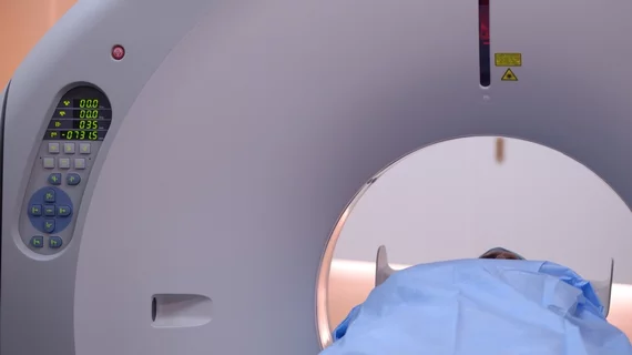
Low-dose contrast-enhanced CT scans are just as effective at diagnosing appendicitis compared to standard scans, according to new research published in the British Journal of Surgery.
Appendicitis is one of the most common ailments seen in emergency departments, and appendectomies are among the most frequently performed surgeries in the world. With non-surgical treatments emerging, such as antibiotics for milder cases, it is important for doctors to accurately differentiate complicated versus uncomplicated appendicitis.
However, this has historically come at the expense of increased radiation exposure for the patient, which is especially concerning for younger individuals. Because of this dilemma, researchers wanted to assess the diagnostic accuracy of low-dose contrast-enhanced CT scans compared to conventional techniques.
Between April 2017 and November 2018, researchers examined 856 patients who presented to three separate emergency departments with suspected appendicitis. Of those patients, 454 underwent LDCT scans and 402 received an exam with standard dosages. The lower doses were calculated using patient BMI, with no one with a number greater than 30 receiving a reduced-dose scan.
The accuracy of low-dose CTs in detecting uncomplicated acute appendicitis was 98%, while the standard dose was just barely higher at 98.5%. These numbers decreased slightly when diagnosing complicated appendicitis but stayed in consistent detection ranges between 90.3% and 87.6%.
The median dose for patients who underwent low-dose CTs was 3 mSv, while the standard scans doubled a patient’s dose to 7 mSv.
“These findings will hopefully encourage physicians to implement low-dose CT modalities at emergency departments for acute appendicitis imaging to avoid unnecessary radiation in this very large patient population,” Paulina Salminen, MD, PhD with the Division of Digestive Surgery and Urology at Turku University Hospital, and co-authors wrote.
You can read the detailed results of the study here.

