New algorithm improves MRI for pregnancy monitoring
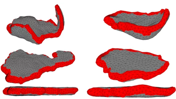
An algorithm designed to unfold MRI scans of the placenta may allow doctors to more accurately identify and treat issues with the organ during pregnancy, according to new research out of MIT’s Computer Science and Artificial Intelligence Laboratory.
A properly functioning placenta is vital to a successful pregnancy. However, traditional imaging techniques such as ultrasound provide limited insight into placental health, according to co-lead author of the research Mazdak Abulnaga. Even more advanced modalities like MRI have trouble imaging the curved surface of the uterus.
“The idea is to unfold the image of the placenta while it’s in the body, so that it looks similar to how doctors are used to seeing it after delivery,” Abulnaga, also a PhD student, added in a news release. “While this is just a first step, we think an approach like this has the potential to become a standard imaging method for radiologists.”
The study is not yet peer-reviewed, but is available online in Cornell University’s repository: arXiv.org. It’s scheduled to be published next month and will be presented Oct. 14 at the International Conference on Medical Image Computing and Computer-Assisted Intervention in Shenzhen, China.
A key aspect of their algorithm is its ability to clearly show cotyledons—“circular structures that allow for the exchange of nutrients between the mother and her developing child.” If radiologists could see these, the researchers wrote, doctors could diagnose and treat placental issue far earlier in the pregnancy.
“The biological processes that underlie cotyledon patterning are not completely understood, nor is it known whether a standard pattern should be expected for a given population,” Chris Kroenke, associate professor at Oregon Health and Science University, who was not part of the study, said in the statement. “The tools provided by this work will certainly aid researchers to address these questions in the future.”
The source code used in this research is available for free on GitHub.

