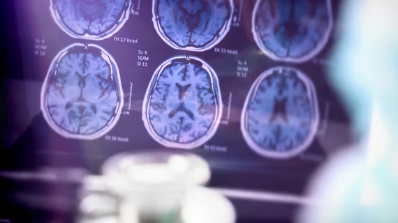
COVID-19 patients with neurological problems requiring brain imaging face increased risk of death
Patients hospitalized with a COVID-19 infection serious enough to require brain imaging face a higher risk of death compared to others, according to a new study published Friday.
Albert Einstein College of Medicine and Montefiore Health System researchers compared nearly 600 patients with neurological issues warranting neuroimaging to more than 1,500 participants with a similarly severe COVID-19 diagnosis but without brain problems.
Those with an altered mental state or imaging-confirmed stroke were more likely to die in the hospital compared to matched controls.
Emergency physicians should consider identifying and treating these individuals to reduce the overall COVID-19 mortality rate, authors explained in Neurology.
"This study is the first to show that the presence of neurological symptoms, particularly stroke and confused or altered thinking, may indicate a more serious course of illness, even when pulmonary problems aren't severe," David Altschul, MD, chief of the Neurovascular Surgery Division at Einstein and Montefiore, said in a statement. "Hospitals can use this knowledge to prioritize treatment and, hopefully, save more lives during this pandemic."
The investigators looked at data from 4,711 COVID-19 patients admitted to Montefiore between March and April 16, 2020. Of that total, 581 (12%) suffered neurological troubles significant enough to require brain imaging. That subset of patients was compared to an age-matched control group of 1,743 individuals with a similar COVID-19 diagnosis.
Among the group who underwent neuroimaging, 55 were diagnosed with stroke and 258 showed confusion or an altered mental state. Those with stroke were twice as likely to die compared to the control group. Inpatients with confusion, meanwhile, had a 40% mortality rate compared to 33% in their matched counterparts.
Of note, Altschul et al. found more than 50% of stroke sufferers did not have hypertension or underlying risk factors for the cerebrovascular accident, a “highly unusual” finding, they added.
Read the entire study published Dec. 18 in Neurology here.

