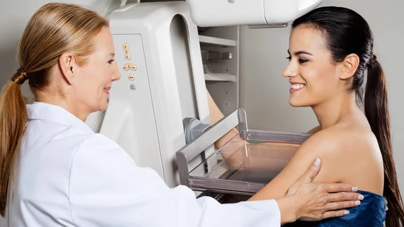
CAD tool detects microcalcification clusters in mammograms
A fully automated algorithm can accurately detect microcalcification clusters in mammogram scans and may help reduce radiology workloads.
Detailed in an August 7 study published in the Journal of Digital Imaging, the computer-aided detection (CAD) tool uses a three-step process and can detect microcalcification (MC) clusters with 100% sensitivity.
Often found in clusters, MCs are small mineral deposits such as calcium that are scattered throughout the breast tissue, explained Vikrant A. Karale, with the Indian Institute of Technology in Kharagpur, India, and colleagues. MC clusters alone are an early indicator of up to half of all non-palpable breast cancers and exist in 93% of ductal carcinoma in situ cases.
“Thus, detection of MC clusters is very crucial in the diagnosis of breast cancer,” the authors added. “Computer-aided diagnosis system(s) can act as a second reader and can help the radiologists to find MC clusters that radiologist otherwise might have missed.”
Karale and colleagues’ CAD algorithm utilized a three-step process to pre-emphasize microcalcifications and detect positive MC clusters, called multiscale 2D NEO; segregate microcalcifications from false positive objects; and finally, address what they described as a data imbalance problem stemming from an uneven number of images containing MCs.
The algorithm’s performance was evaluated on three databases and went through a five-fold validation process. Overall, the algorithm achieved a 100% sensitivity with false positive rates per image of 2.59, 1.78 and 0.68 on each of the three databases.
However, the authors noted, the multiscale 2D NEO failed to achieve a 100% sensitivity for detecting individual microcalcifications.
Despite this, the team believes their CAD platform could be used as a screening tool and reduce the ever-growing workload many radiologists face, especially in African countries where readers are often in short supply.
“Apart from the normal use of CAD as a second reader, the mean multiscale 2D NEO can act as a screening tool,” the group concluded. “It might eliminate considerable portion of the normal images from the work-list and thereby reduces the workload of the radiologists.”

