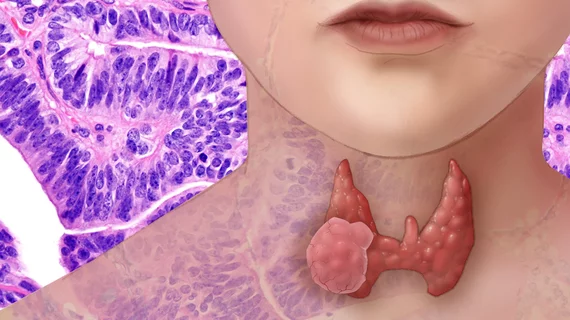
New framework pushes radiology toward fully automated triage system for thyroid cancer
A new framework can predict if a thyroid nodule is cancerous similarly to a trained expert. And with further development, the technique could usher in an automated system for triaging such nodules.
Stanford University researchers described their approach—which extracts quantitative features such as texture and edge sharpness from thyroid biopsies to predict their malignancy—Jan. 22 in the American Journal of Roentgenology. The model performed better than a number of human classifiers and, with more work, could make thyroid cancer care much more efficient.
“Once the algorithms and framework are devised, a fully automated process of thyroid nodule triage would be within reach,” Alfiia Galimzianova, with Stanford’s Department of Biomedical Data Science, and colleagues wrote. “Development of an automated image analysis system would be the first step toward implementation of large-scale automated screening of thyroid image datasets,” they added.
Diagnosing thyroid nodules via imaging findings remains a challenge. Currently, this can only be accomplished with tissue biopsy or surgery, but even then, only an estimated 5%-7% of nodules are ultimately considered malignant. As a result, “most” patients are exposed to unnecessary health risks related to testing, pushing healthcare costs ever-higher, the team wrote. There is a “critical” need to stratify the risk of thyroid nodules and “intensive” research has been underway in radiology and other specialties to find an answer.
With this in mind, the team tasked their “elastic net classifier” to diagnose an ultrasound dataset of 92 biopsy-confirmed nodules. Two expert radiologists annotated the images using the thyroid imaging reporting and data system.
“By extracting a rich set of features from these images in a computerized manner, our proposed framework expands the scope of features for analysis beyond merely those that are visible to the human eye and thereby maximizes use of the information available in the images,” Galimzianova et al. wrote. “The use of computer-aided diagnostic tools based on frameworks similar to ours could improve the management of thyroid nodules and decrease the number of unnecessary biopsies and surgical risk while care is more appropriately directed at patients who need more invasive management.”
One key limitation of this study is that the approach still requires a radiologist to manually segment nodules, a tedious and time-consuming task. Going forward, however, the team will look to create an algorithm to complete this process, they concluded.

