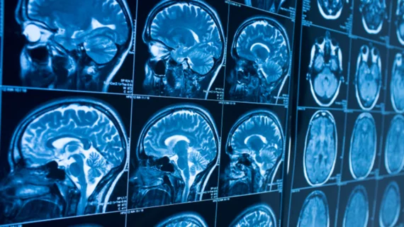
Brain tumor reporting system improves quality of glioma reports
Radiology reports derived from structured brain tumor MRI reporting and data systems (BT-RADS) showed measurable improvements compared to free text reports, according to a new study published in Academic Radiology.
A multidisciplinary group, including James Y. Zhang, MD, PhD, with Emory University School of Medicine in Atlanta, compared nearly 400 glioma reports before and after implementing BT-RADS. They found quantitative improvements in many reporting features including historical elements such as chemotherapy and radiation history—central to brain tumor treatment.
“As compared to conventional free prose reports, reports derived from BT-RADS demonstrated superior traits across multiple domains, including greater consistency and completeness of history reporting, reduced ambiguity, and improved conciseness,” Zhang and colleagues wrote.
BT-RADS was developed for classifying and reporting post-treatment brain tumor images, particularly gliomas. These tumors are “intrinsically challenging for reporting,” the authors noted, due to the vast amounts of clinical information for each patient and the “heterogenous nature” of cases.
“Because of the subjectivity of conventional free prose reporting, this can introduce variability, inconsistency, and ultimately confusion in reporting,” the researchers added. “This can lead to miscommunication between the radiologist and other members of the healthcare team, and even between different radiologists who may be interpreting the same images at varying time points.”
The researchers administered a survey prior to, and after implementing BT-RADS to gauge clinician’s opinions on the quality of glioma reports. Three complaints were most prevalent: unclear communication, inconsistent reporting, and too much ambiguity.
Zhang and colleagues then analyzed 211 pre-BT-RADS and 172 post-BT-RADS reports, assessing clinical history text, the usage of hedge words to measure ambiguity, report length and the use of addendums.
Reports generated after implanting BT-RADS included more history words, including “Avastin” (7.6% vs. 20.9%) and methylguanine-DNA methyltransferase (10.9% vs. 31.4%). BT-RADS reports also included less hedge words such as “possibly” and “likely.” Newly generated reports were also shorter and contained fewer addenda.
“Given these findings, BT-RADS is a promising new structured report system that can benefit any institution performing imaging following glioma treatments,” the researchers wrote.
There was not much emphasis placed on analyzing the subtle variations and nuances of language in the reports, the authors noted, which may have offered some benefit in free prose reports. Despite this, Zhang et al. thinks the future is bright for BT-RADS.
“Structured report systems remain an evolving field, with many potential areas for improvement and opportunities for synergy,” Zhang and colleagues wrote. “One particularly promising synergistic partner is artificial intelligence, which is already gaining much traction among radiologists and has tremendous potential.
Thus, this may emerge as another area of innovation for BT-RADS.”

