High-strength MRI may release mercury from silver fillings
A new study, published online June 26 in Radiology, found ultra-high strength MRI may release toxic mercury from silver fillings, though commonly used 1.5 Tesla MRI machines appear to have no effect on the cavity-prone.
Amalgam fillings, commonly called silver fillings, are nearly 50 percent mercury—but the U.S. Food and Drug Administration (FDA) still considers them safe for adults and children over 6 years old, according to a Radiological Society of North America (RSNA) release.
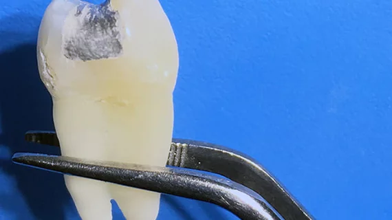
Selmi Yilmaz, PhD, and faculty dentist at Akdeniz University in Turkey, and Mehmet Zahit Adişen, PhD, with the faculty of dentistry at Turkey’s Kirikkale University, examined 60 teeth extracted for clinical indication.
The two opened two-sided cavities in each tooth and inserted amalgam fillings. Nine days later, 20 randomly selected teeth were split into two groups—one was placed into a solution of artificial saliva and exposed to 1.5-T or 7-T MRI for 20 minutes, and the other was placed only in the saliva as a control.
"[W]e found very high values of mercury after ultra-high-field MRI," Yilmaz said in the RSNA statement. "This is possibly caused by phase change in amalgam material or by formation of microcircuits, which leads to electrochemical corrosion, induced by the magnetic field."
The use of 7-T MRI was approved by the FDA in 2017, but its availability is extremely limited, according to the release.
Additionally, authors noted “it is not clear how much of this released mercury is absorbed by the body.” They suggested further studies may be needed to determine the relationship between high-field MRI and the release of mercury from amalgam fillings.
Patients with silver fillings should have no concern about having a 1.5-T MRI exam, the release stated.

