Back to school: What radiologists can learn from bipedal evolution
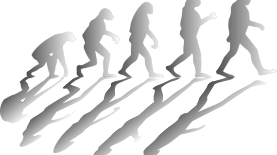
The ability to walk upright on two feet required millions of years of evolution, and even today humans are not perfectly adapted to bipedalism, argued authors in a recent Academic Radiology perspective.
Many complaints that require routine imaging can be traced to this evolutionary change, prompting the authors from Indiana University School of Medicine in Indianapolis to ask: How did bipedalism evolve? And what challenges does it present for spine, pelvis and lower limb function?
“Gaining a deeper understanding of these questions can help radiologists better understand the genesis of a diverse range of conditions we encounter on a daily basis, such as traumatic and insufficiency fractures, osteoarthritis, spondylolisthesis and even disorders with no apparent relationship to upright posture,” the pair wrote.
Richard B. Gunderman MD, PhD, and Abdul R.S. Asar provided the human spine as a prime example. It originally developed parallel to the ground, but eventually gave way to its signature curve—enabling balance, but increasing risks.
Intervertebral disc injuries and forms of misalignment are all exclusively human problems, the authors wrote. The foot, once meant for grasping, is also at an increased risk for conditions such as tendonitis and arthritides due to extra weight bearing needs.
Around the time hominids began walking upright, the hips and knees began to change, which in turn altered the way the knee works—making it prone to osteoarthritic change, Gunderman et al. argued.
Pair these problems alongside a lifespan longer than most mammals, and humans become more vulnerable to overuse and degenerative diseases.
Maintaining a healthy weight and practicing regular weight-bearing exercises can reduce osteoporosis and strengthen supporting bones and joints, the authors wrote.
“While understanding these incomplete biological adaptations offers little as a means of enhancing diagnostic acumen, it does help to make sense of a large range of otherwise seemingly unrelated diseases and injuries that radiologists encounter on a daily basis,” Gunderman and Asar concluded.

