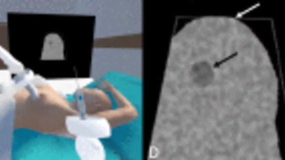
VR could improve breast biopsy skills for trainees
Virtual reality training could have great utility for improving residents’ proficiency in completing breast biopsy procedures.
Breast biopsies are the most common image-guided procedure. Despite this, many radiology residents may not be routinely involved in completing them during their training. Given the importance and frequency of the procedure, it is vital that students gain competence in completing it.
Current simulation training provides more chances for students to practice the skill, but the emergence of VR could present an opportunity to improve training with a more realistic environment, authors of a new paper in Current Problems in Diagnostic Radiology propose.
“Current simulation training often involves the use of manufactured or homemade (chicken or turkey breast) phantoms. Virtual reality is an emerging technology, allowing learners to have flexibility in learning, real-life interactive experiences and measurable feedback,” corresponding author Stefanie Woodard, with the Department of Radiology at the University of Alabama at Birmingham, and co-authors explain. “A fully immersive VR simulation would allow a trainee to have an opportunity to enter a breast biopsy room with the patient, instruments and ultrasound machine.”
Researchers sought to determine whether VR could be a feasible training option for conducting ultrasound-guided breast biopsies. To do this, they had three fellowship-trained breast radiologists test a VR breast biopsy simulation program. The group first underwent a brief training session describing how to navigate throughout the simulation, before being instructed to conduct as many biopsies as possible within 15 minutes. Time to successful biopsy, body entries, and users’ self-reported experiences were recorded.
The simulation was well tolerated and did not trigger motion sickness or fatigue in any of the participants. The radiologists’ time to successful biopsy significantly reduced with each subsequent attempt after their initial simulated procedure completion. The VR simulation also reduced the group’s amount of body entries and biopsy fires required to acquire a successful tissue sample.
“As with an actual biopsy, once the spatial alignment of the probe, needle, and lesion is determined, repeated lesion sampling typically takes less time,” the group notes. “The biopsy simulation also demonstrates this, suggesting the simulator allows for a reproducible biopsy target and muscle recognition of spatial alignment.”
All three radiologists gave the simulation training high marks. The group found it to be engaging and realistic, noting that they would recommend it to trainees as well.
Future testing with residents and medical students is in the works.

