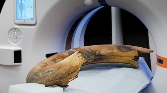
A mammoth accomplishment: CT captures images of an entire ancient tusk
Experts were recently able to visualize an entire woolly mammoth tusk with the help of a large CT scanner—a feat that was accomplished in one fell swoop for the first time.
Prior attempts at imaging large fossils such as mammoth tusks failed to capture the full artifact with just one scan, instead requiring multiple partial scans that were subsequently pieced together. A new “Images in Radiology” article published recently in Radiology details how experts were able to achieve this marked accomplishment to estimate the mammoth’s age thanks to the help of newer CT technology.
“Working with precious fossils is a challenge since it is important not to destroy or harm the specimen,” said the article’s senior author, Tilo Niemann, MD, head of cardiac and thoracic radiology in the Department of Radiology at Kantonsspital Baden in Switzerland. “Even if there exist various imaging techniques to evaluate the internal structure, it was not possible to scan a whole tusk in toto without the need for fragmentation or at least having to do multiple scans that then had to be painstakingly assembled.”
Woolly mammoths were similar in size to modern day African elephants. This tusk was measured at nearly seven feet long, with a total object diameter of just over 2.5 feet, so the analysis required a CT gantry large enough to accommodate its immense size.
The CT images produced detailed depictions of the tusks “cones,” which are actually annual increments of dentin apposition that resemble cone-shaped cups and are used to estimate the age of mammoths. Using the CT images, the experts observed a total of 32 cones, suggesting that the mammoth was around 32 years old at the time of its death, which likely occurred close to 17,000 years ago.
View more of the acquired images here.

