Computed Tomography

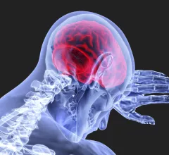
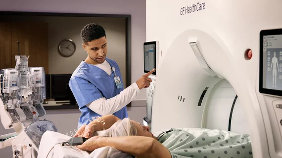
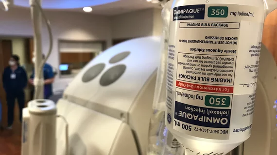
Computed tomography (CT) is a fast and accurate imaging modality often used in emergency settings and trauma imaging. CT scans, with or without (or both) iodinated contrast are frequently used to image the brain, chest, abdomen and pelvis, but also have post-imaging reconstructive capabilities for detailed orthopedic imaging. It is now a standard imaging modality in emergency rooms to quickly assess patients. CT uses a series of X-ray images shot as the gantry rotates around the patient. Computer technology assembles these into into a dataset volume than can be slices on any access, or advanced visualization software can extract specific parts of the anatomy for study. Find more content specific to cardiac CT.
Thumbnail

Offering imaging exam for free ups utilization by 546%, with key gains in underserved populations

University Hospitals Health System tested the pricing change in a bid to boost outcomes in its service area. 

Thumbnail

How Secure Is That Scanner?

In a world of networked medical devices, it’s not hard to imagine a radiology-heavy cyberattack that is not only malicious but also ingenious.
 

Thumbnail

Plaque characteristics boost predictive power of CTA risk scoring

A CT angiography (CTA)-derived score that also incorporated the extent, location and composition of coronary plaque outperformed a model that focused only on the severity of stenosis, researchers reported Jan. 16 in JACC: Cardiovascular Imaging.

Thumbnail

Industry group releases new guidelines for CT use in TAVR procedures

The Society of Cardiovascular Computed Tomography (SCCT) released updated guidelines for using CT imaging in patients undergoing transcatheter aortic valve replacement (TAVR).

Fujifilm announces entry Into U.S. computed tomography market with unveiling of FCT Embrace at ASTRO 2018

World's first 85cm 64 and 128 slice wide bore CT unit for oncology

Example of spectral, or dual-energy CT, confirming a pulmonary embolism (PE). Image courtesy of Philips Healthcare

Dual-energy CTA iodine maps offer small benefit in diagnosing pulmonary embolism

Dual-energy (DE) CT iodine maps offer a slight benefit when paired with traditional CT angiography images in diagnosing pulmonary embolism (PE), reported researchers in a Sept. 11 Radiology study.

Perivascular fat attenuation index CT scans showing the amount coronary wall inflammation. This measure is being tested in trials to see if it can accurately predict which coronary artery lesions will progress to cause heart attacks.

Novel cardiac imaging biomarker could predict risk of coronary inflammation, heart attack

A team of international researchers has developed a new imaging biomarker able to non-invasively predict a patient’s risk of coronary inflammation and heart attack, according to research published Aug. 28 in The Lancet.

Thumbnail

Document offers guidance on curbing radiation exposure during CV imaging

An expert consensus document published May 2 in the Journal of the American College of Cardiology offers recommendations for limiting radiation exposure to both patients and medical personnel during cardiovascular imaging procedures.

Around the web

The nuclear imaging isotope shortage of molybdenum-99 may be over now that the sidelined reactor is restarting. ASNC's president says PET and new SPECT technologies helped cardiac imaging labs better weather the storm.

CMS has more than doubled the CCTA payment rate from $175 to $357.13. The move, expected to have a significant impact on the utilization of cardiac CT, received immediate praise from imaging specialists.

The newly cleared offering, AutoChamber, was designed with opportunistic screening in mind. It can evaluate many different kinds of CT images, including those originally gathered to screen patients for lung cancer. 

Trimed Popup
Trimed Popup