Computed Tomography

Computed tomography (CT) is a fast and accurate imaging modality often used in emergency settings and trauma imaging. CT scans, with or without (or both) iodinated contrast are frequently used to image the brain, chest, abdomen and pelvis, but also have post-imaging reconstructive capabilities for detailed orthopedic imaging. It is now a standard imaging modality in emergency rooms to quickly assess patients. CT uses a series of X-ray images shot as the gantry rotates around the patient. Computer technology assembles these into into a dataset volume than can be slices on any access, or advanced visualization software can extract specific parts of the anatomy for study. Find more content specific to cardiac CT.
Treatment costs of CT-related cancers could reach $2.6 billion over 10 years

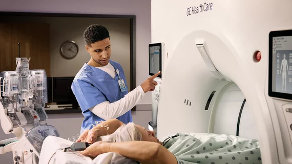
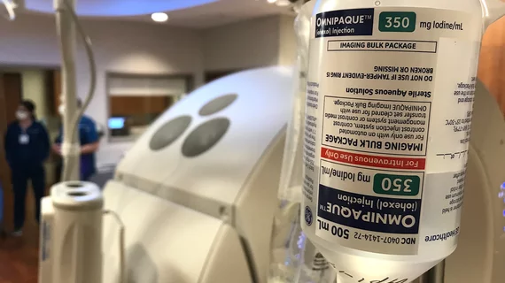
Despite the many benefits of CT imaging—early diagnosis, enhanced diagnostic accuracy, improved outcomes, etc.—a small number of cancers resulting from patient exposure to radiation from CT scans can have a big financial impact on treatment costs, according to results of a study recently presented at the annual meeting of the American Roentgen Ray Society.

Budoff wins SCCT’s 2016 gold medal award

The Society of Cardiovascular Computed Tomography (SCCT) announced that Matthew J. Budoff, MD, had won its 2016 Gold Medal Award.

Thumbnail

CT-based Mobile Stroke Care

Germany's ground-breaking use of CT scanners in ambulances to assess for possible stroke inspired one program in Houston to follow suit. Supporters predict it will transform care in the future. 

Radiology: Decision support reins in CT pulmonary angiographic exams

Evidence-based decision support programs may be able to reduce the number of unnecessary CT pulmonary angiographic exams used to evaluate pulmonary emboli in the emergency department, according to a study published online Dec. 20 in Radiology.

Fine Line: Gender Disparities in Cardiac Disease Management

Imaging often the bedrock for first diagnosing and then treating cardiac disease remains challenging and inconsistent in women.

Example of a curved MPR image reconstruction of entire length of of a coronary artery on a cardiac CT scan to better show calcified and soft plaque burden inside the vessel. The thumbnail dots on the left side of the image are cross sectional views of the vessel. Siemens example on the expo floor.

The CCTA Playbook: A Guide to Coding, Reimbursement and Operations

A compendium of the business intelligence required to launch a CCTA service

Around the web

Positron, a New York-based nuclear imaging company, will now provide Upbeat Cardiology Solutions with advanced PET/CT systems and services. 

The nuclear imaging isotope shortage of molybdenum-99 may be over now that the sidelined reactor is restarting. ASNC's president says PET and new SPECT technologies helped cardiac imaging labs better weather the storm.

CMS has more than doubled the CCTA payment rate from $175 to $357.13. The move, expected to have a significant impact on the utilization of cardiac CT, received immediate praise from imaging specialists.