
PHOTO GALLERY: What does fetal medical imaging look like?
This is a clinical photo gallery of fetal imaging that explains what all can be seen on medical imaging, including baby ultrasound imaging, how to determine the sex of a fetus, fetal echocardiograms, how fetal ultrasound measurements are used to track the development of a baby, and how potential issues with a baby can be diagnosed before birth. It includes examples of fetal ultrasound, the primary imaging modality used during gestation, magnetic resonance imaging (MRI) and some rare images of fetal computed tomography (CT).
Hover over images to see the captions. You also can click on images to enlarge them.
A 3D ultrasound fetal image showing the umbilical cord inside the amniotic sac. Image courtesy of Alpinion.
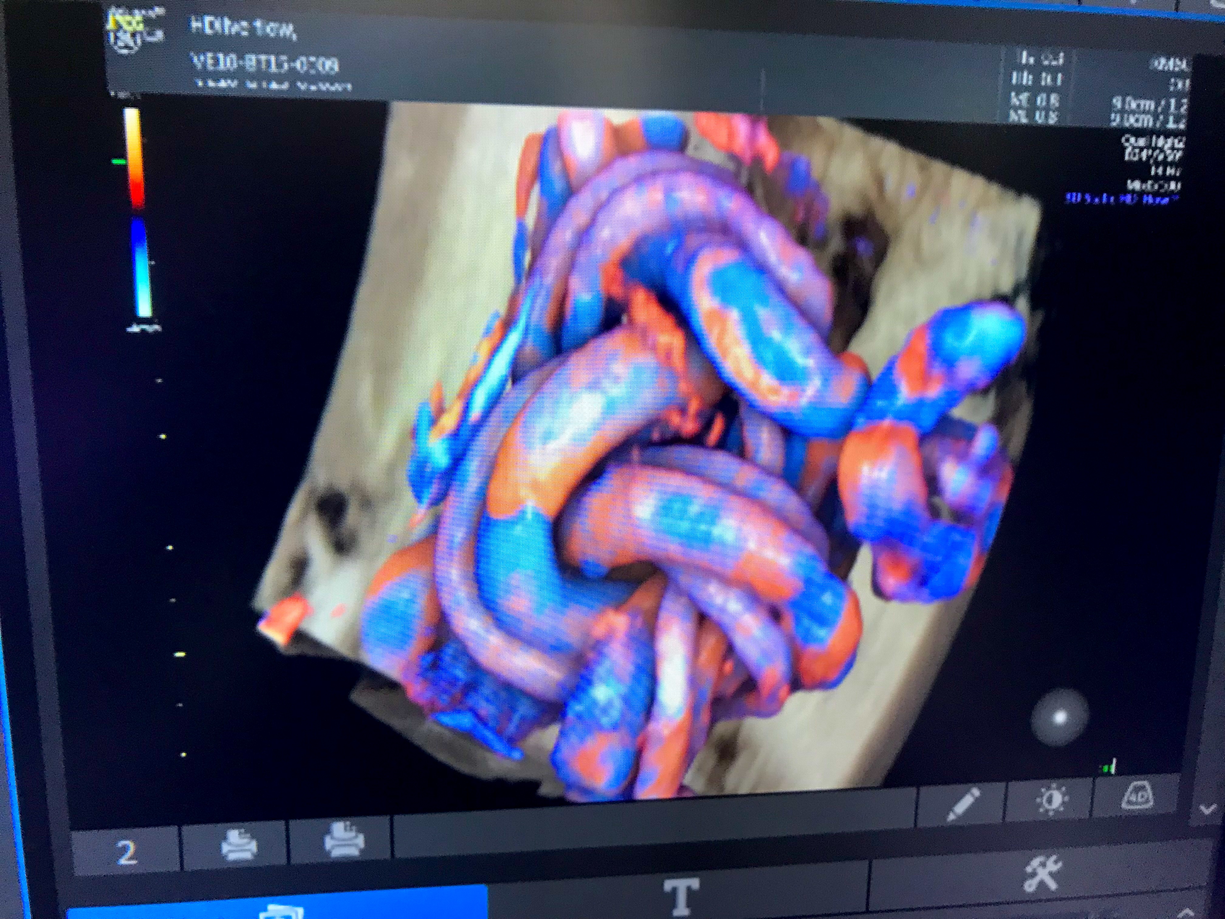
Umbilical cord tied into a knot from fetal movement seen on a 3D/4D ultrasound. The color coding shows the blood flow, which can help determine if the fetus is getting enough oxygen from the mother's blood supply.
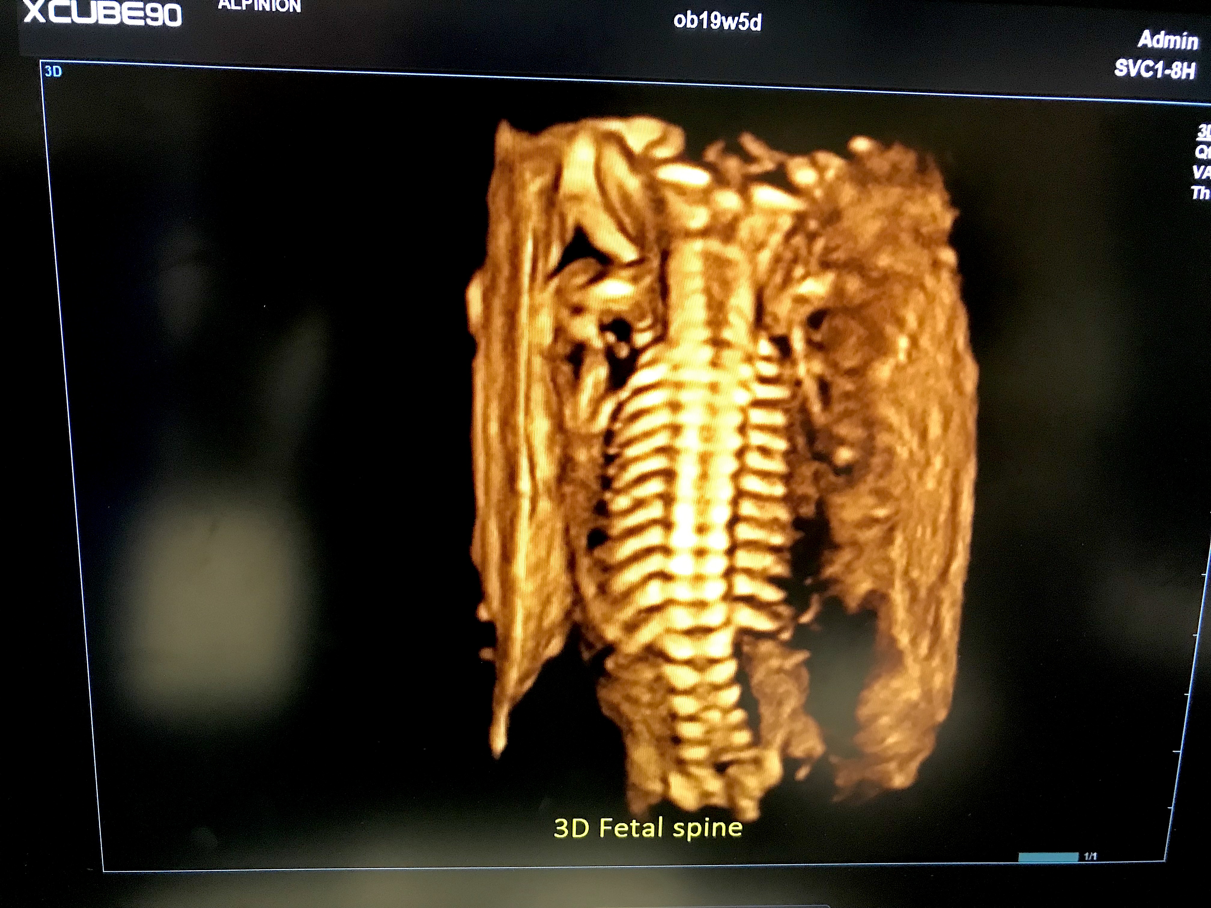
Fetal spine on 3D ultrasound. Image from Alpinion at RSNA 2022.
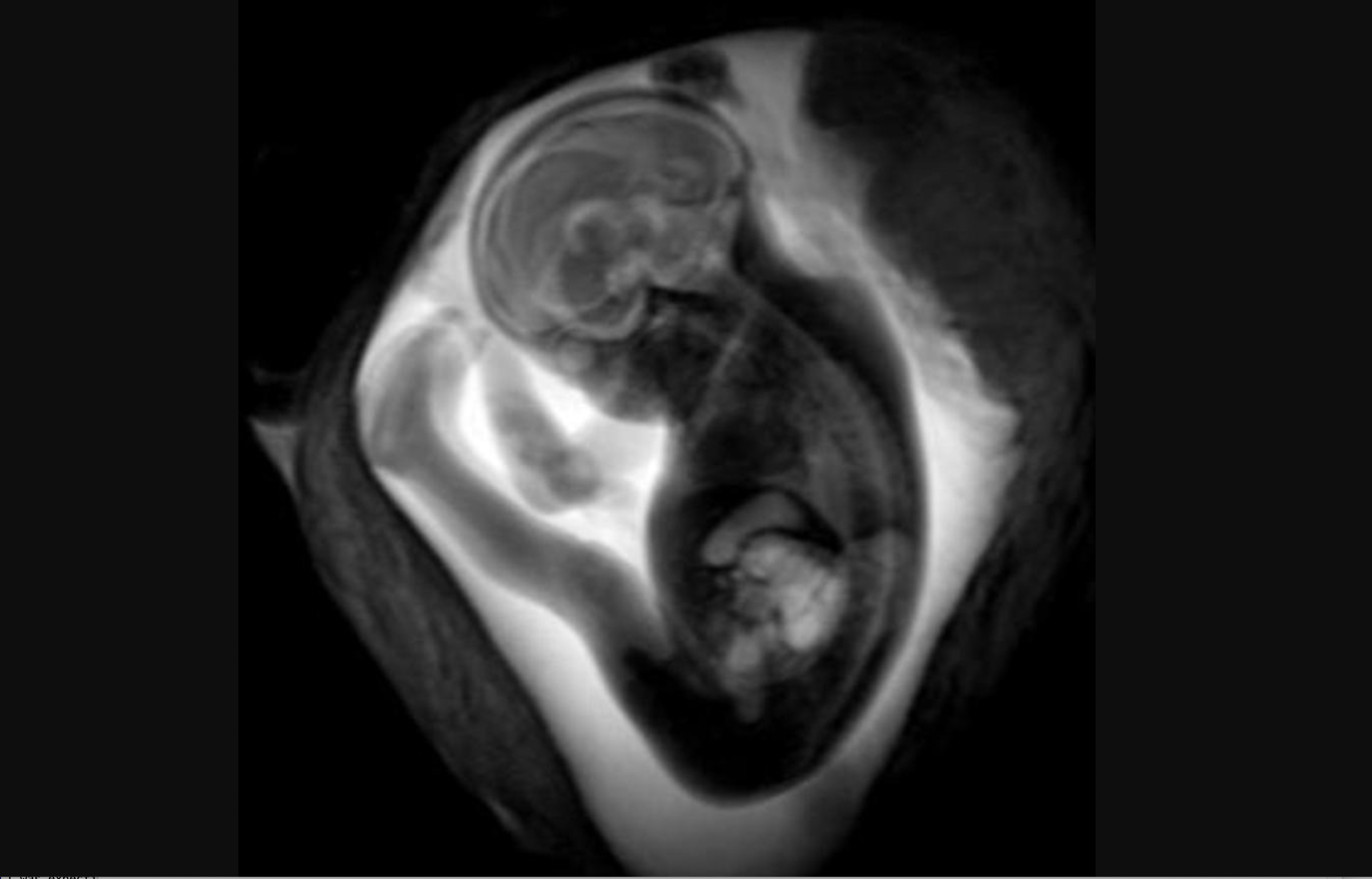
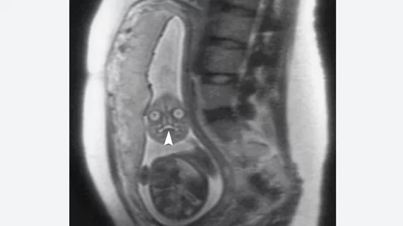
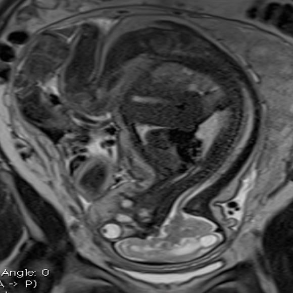
MRI of a fetus inside the mother. Image courtesy of RSNA.
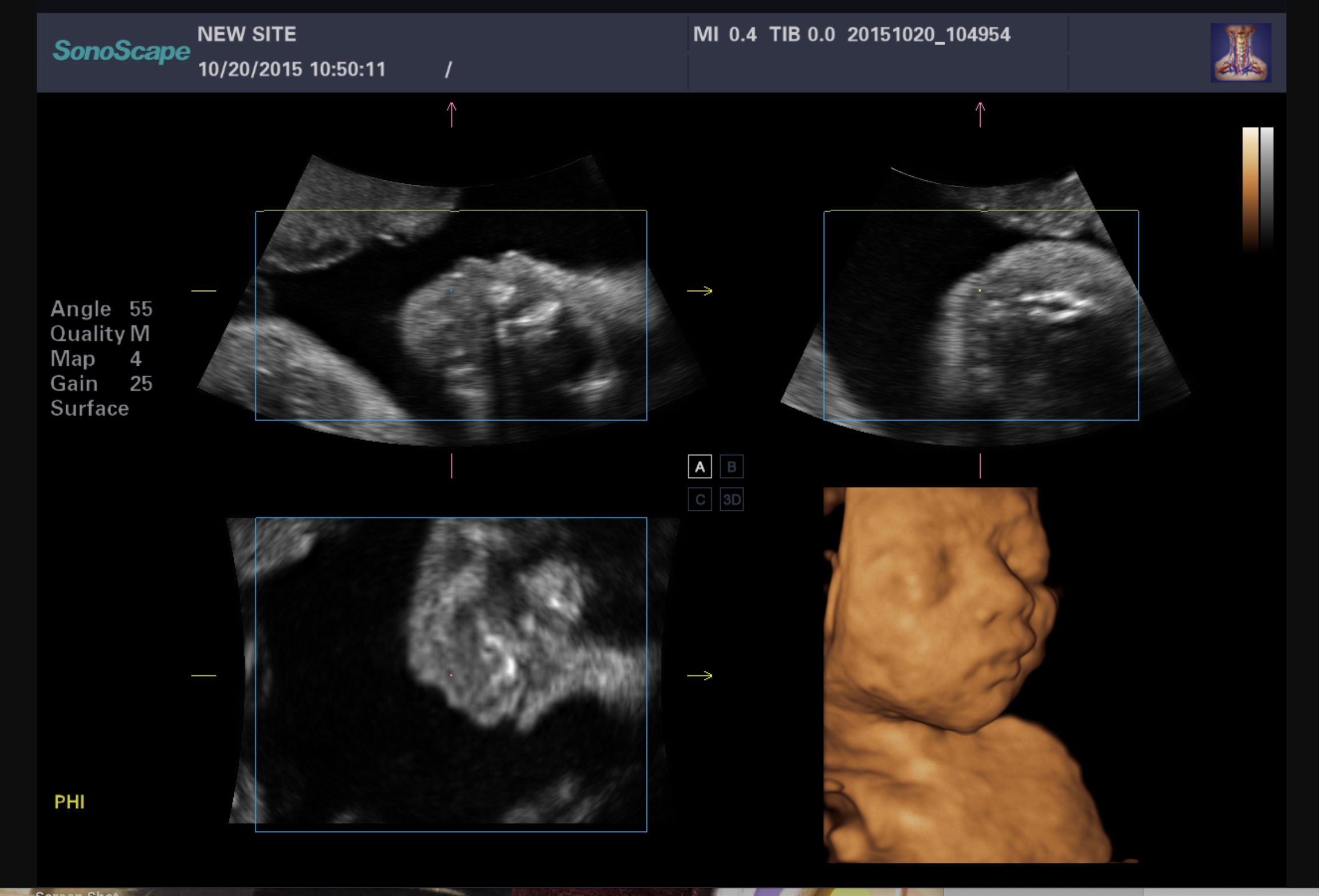
A standard view of a 3D fetal ultrasound exam, where 2D images are shown in the three standard imaging planes, each showing the same anatomy from a different angle, and a rendered 3D volume image. Sonographers and radiologists use the 2D images to make diagnoses and to take measurements. Image courtesy of Konica-Minolta.
Transvaginal ultrasound of a fetus in early development during the first trimester. Image courtesy of RSNA

A patient undergoing a fetal ultrasound exam.
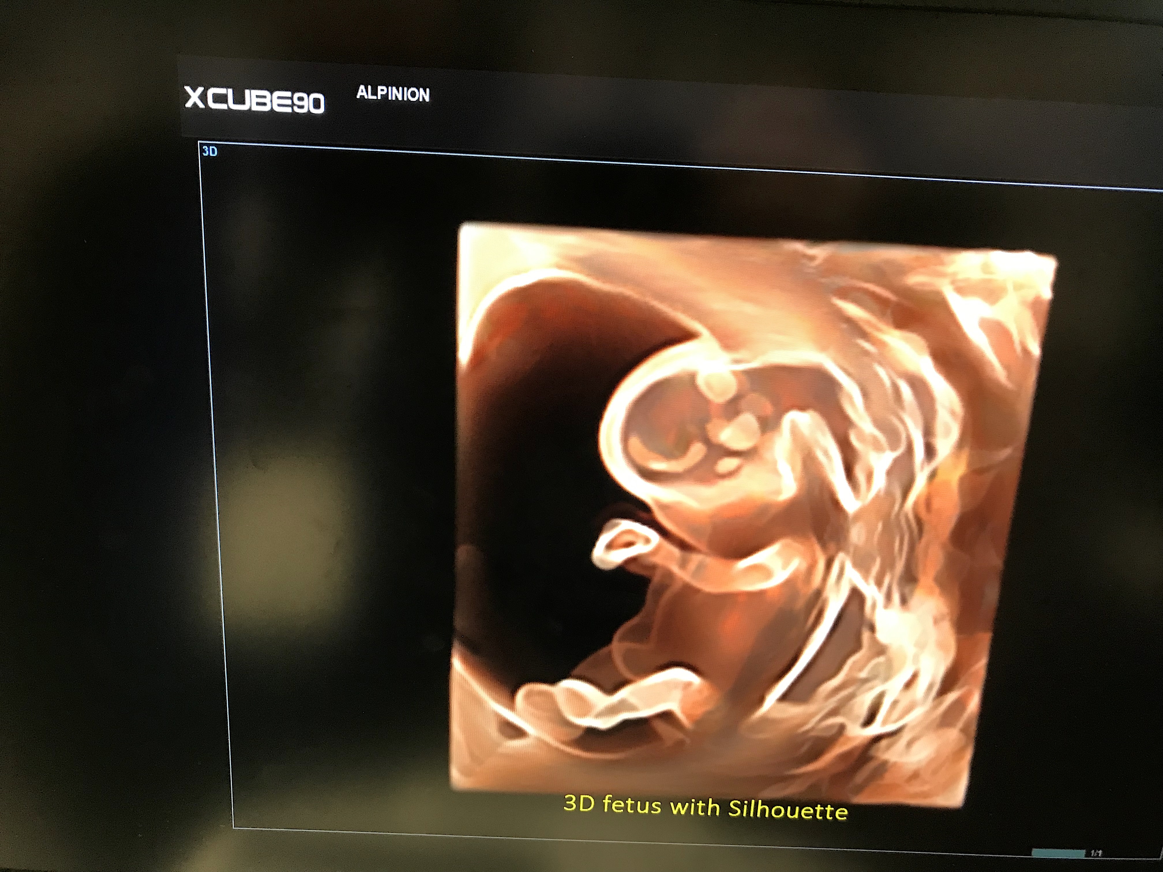
Fetal 3D ultrasound face shown by the vendor Alpinion. The company also OEMs its technology to some of the bigger vendors on the floor at the large RSNA radiology conference.

A 4D fetal heart MRI showing the major vessels and blood flow circulation being developed by the School of Biomedical Engineering and Imaging Sciences, Kings College London. With further development, the method could become a new tool for aiding diagnosis of congenital heart disease where conventional methods like ultrasound might fail. Fetal hearts are so small and beat so fast, it is very difficult to capture details. Rather than just getting a snapshot of the heart, a series of images is taken, allowing cardiologists to see the heart contracting and beating. Image courtesy of King’s College London. Read more
MRI image to assess fetal brain development. Image courtesy of RSNA
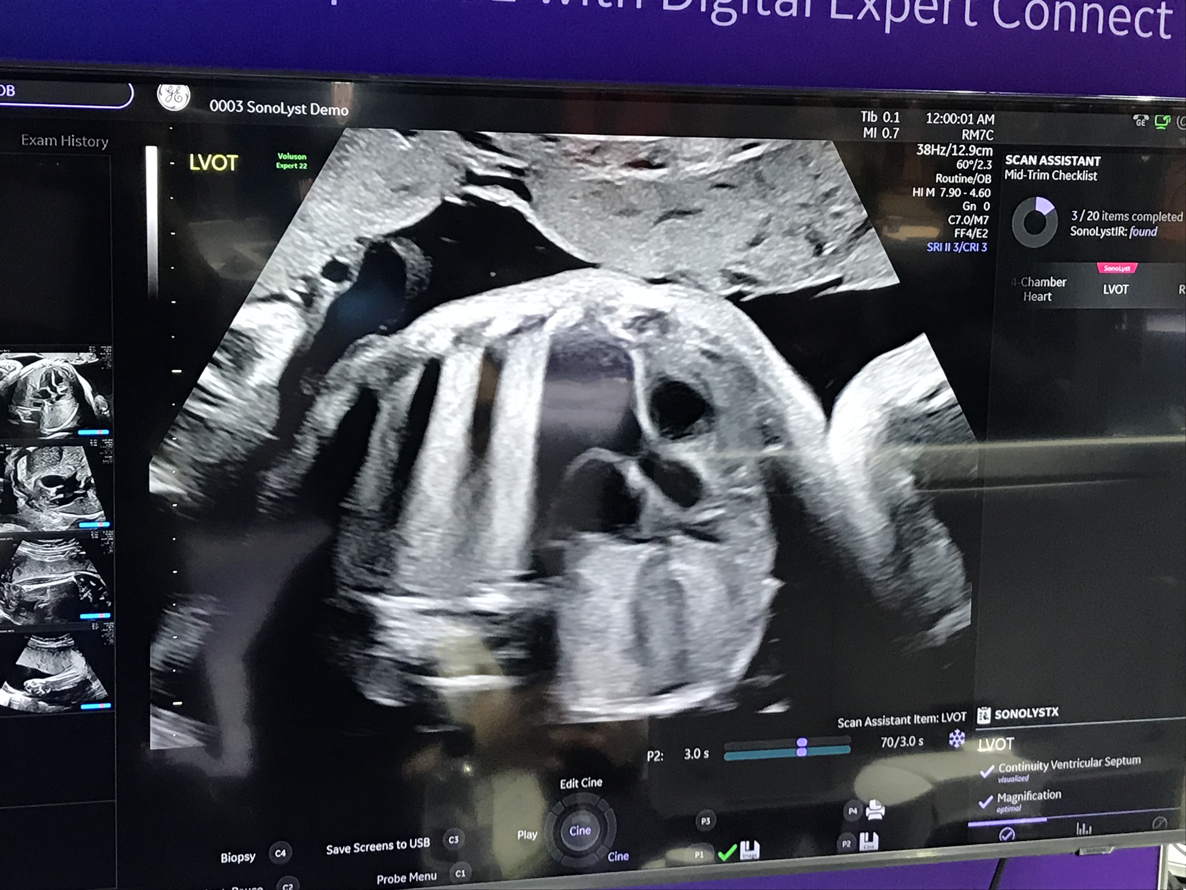
Assessment of the left ventricular outflow tract (LVOT) of the heart and aorta to look for congenital heart defect anomalies. A demo image from GE Healthcare at RSNA 2022.
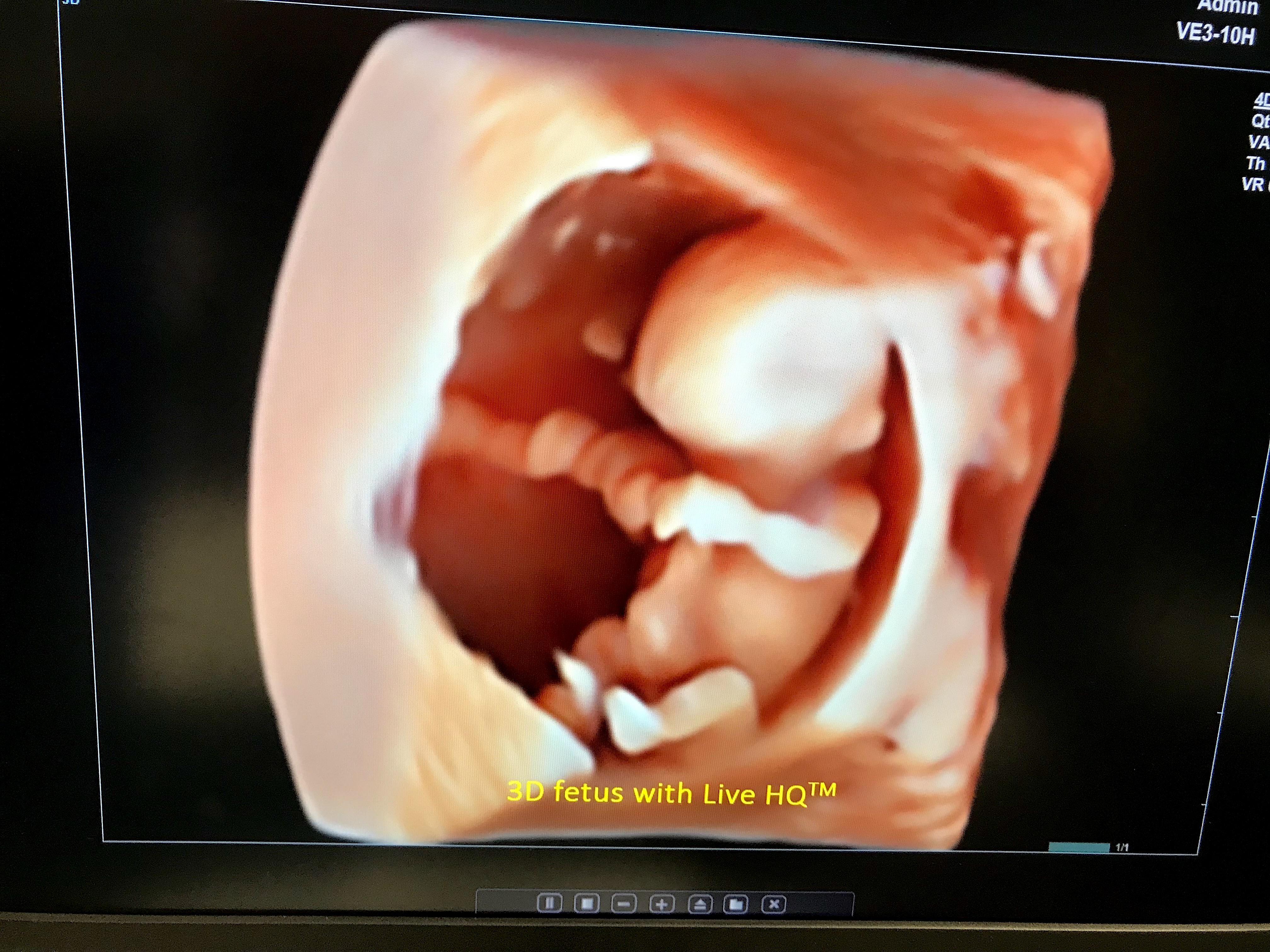
Example of 3D photo-realistic ultrasound technology at RSNA 2022. The simulated lighting source can be moved around to change the locations of the shadows. This makes for great portrait shots for the parents, but also allows physicians to better understand the relationship of anatomical structures if needed during fetal assessments. This is the HDlive rendering technology on the GE Voluson E10 system.
Fetal heart blood flow seen in a Doppler flow 2D ultrasound, showing a regurgitant jet of blood (area of bright yellow) flowing back into a heart chamber due to an issue with a heart valve not sealing properly during systole. This type of assessment can help determine if there are issues with the heart, including congenital heart detects, that will need to be addressed with early surgical interventions after birth. Image courtesy of Alpinion
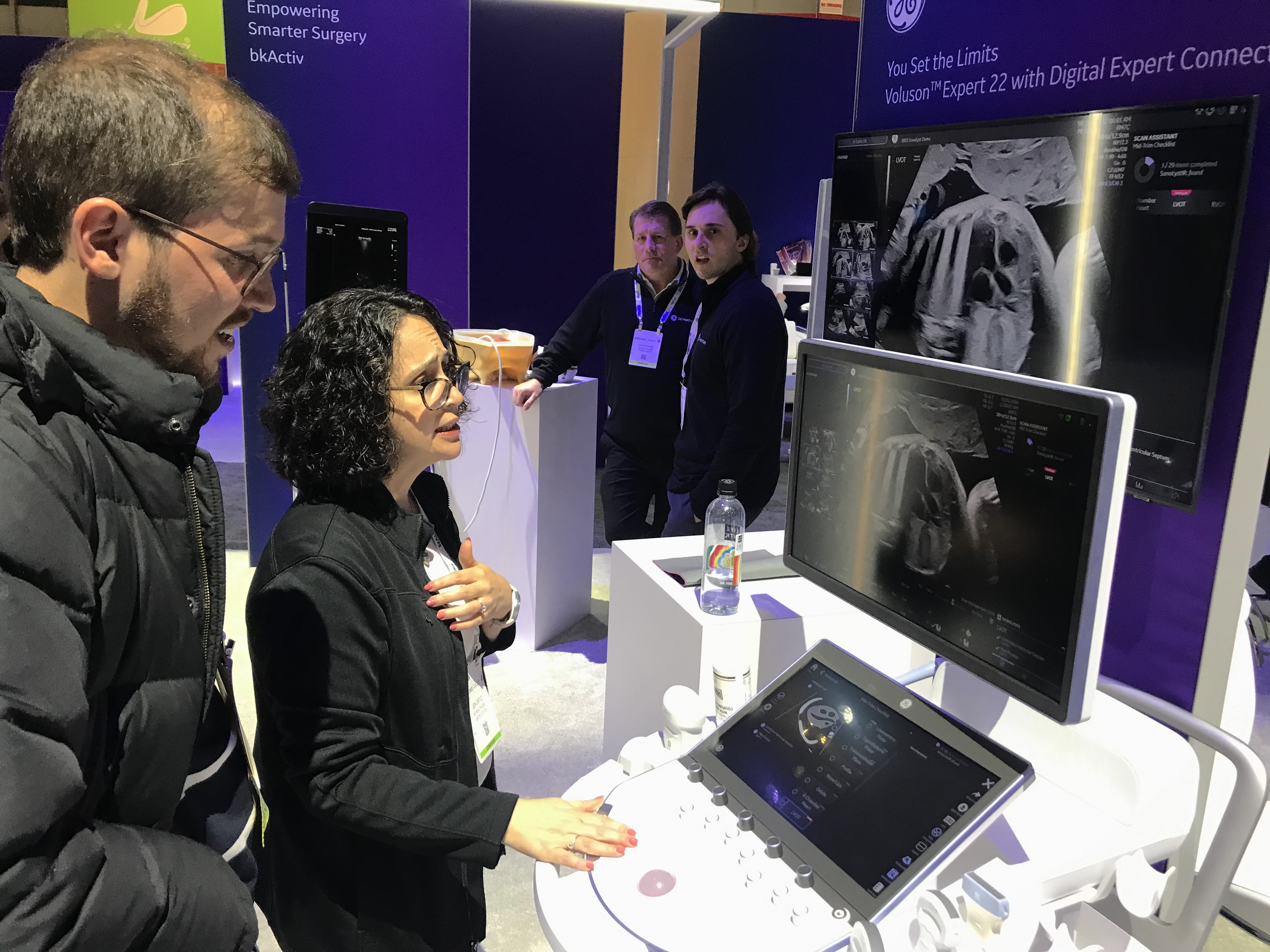
Questions by the sonograpger or radiologist often come up during an exam, especially when there are atypical findings, which often requires a consult with another physician. This is a demonstration at the RSNA 2022 conference for GE Healthcare's Digital Expert technology, showing how users have the ability to interact with peers at various locations to get support before, after, or even during an exam using the GE software. The demo shows a fetal heart and great vessel assessment.
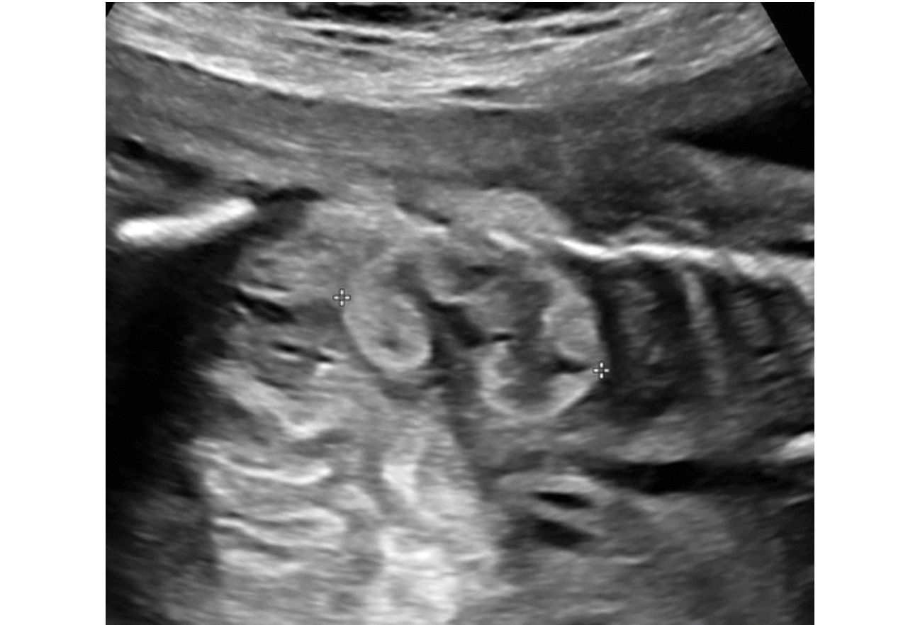
Fetal arm, showing the ulna and radius bones and elbow. Image courtesy of Alpinion
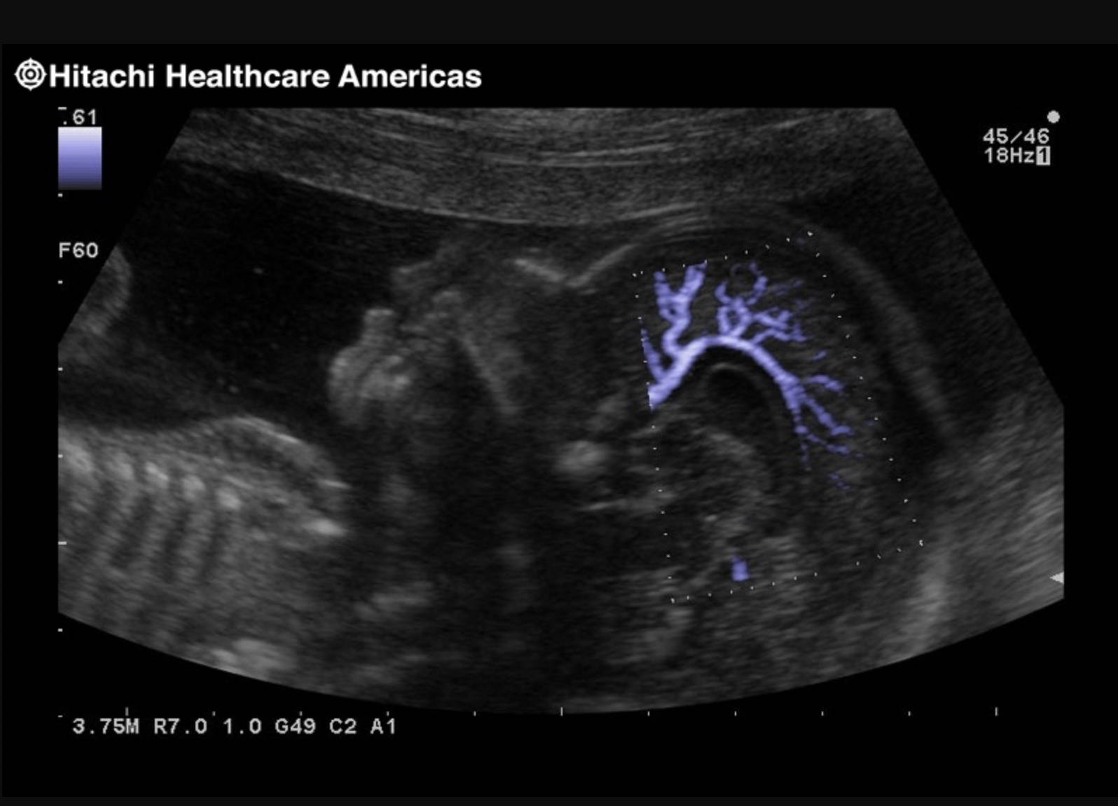
Middle cerebral artery blood flow assessment in the fetal brain. This is used to assess blood flow and fetal hypoxemia. The white oval outline in the images is the fetal skull. Image courtesy of Alpinion.
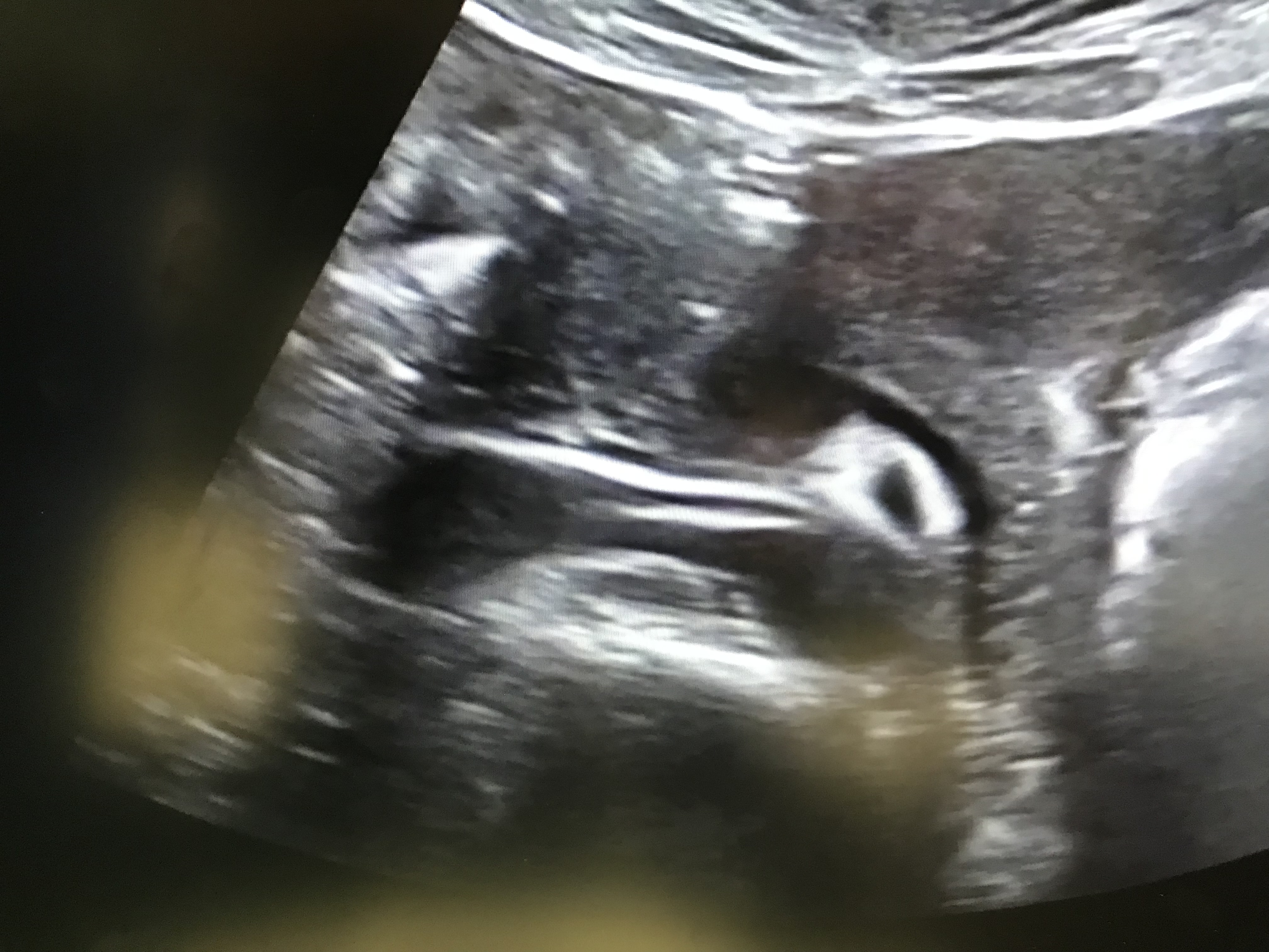
Example of a fetal head profile on 2D ultrasound. This is a common image shown to the mother during exams because it is easy for most people to identify it as a baby, and it shows the outline of the fetal face with the chin and nose. The top of the screen shows the layers of skin, fat and muscle tissue closest to the ultrasound transducer. The gray layer below is the wall of the uterus. The black areas around the face are the amniotic fluid. The white lines that outline the head is the skull and bone starting to form in the bones that make up the skull under the face. The dark shadows that radiate down below the chin and face are acoustic shadows caused by the sound waves not penetrating the areas of bone. Image from Alpinion.
3D ultrasound of a fetus early in development. Image courtesy of Alpinion.
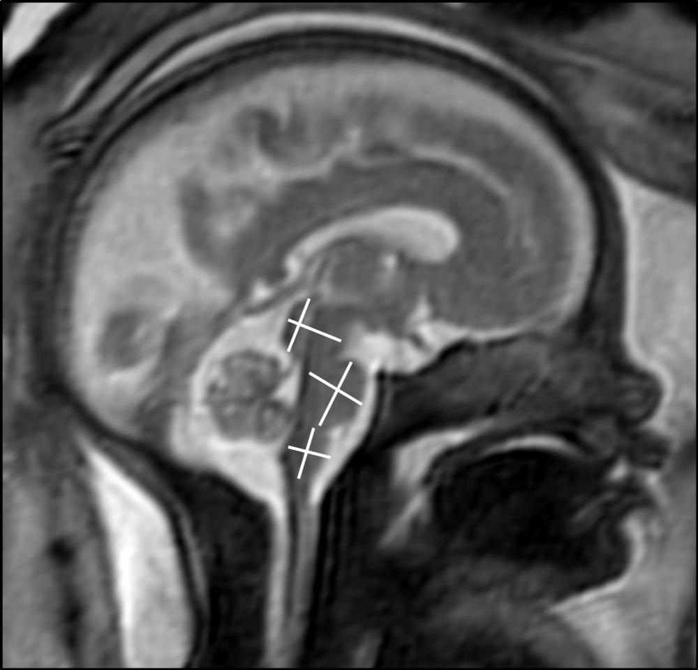
Example of one of the four standard views used in prenatal ultrasound assessment of the head, imaged from the top of the skull looking down into the brain. This is the posterior fossa or transcerebellar view. It is an axial image of a fetus at about 18 weeks gestation at the level of the thalamus or midbrain.
MRI of a fetus infected with zika virus microcephaly, which causes a smaller head circumference measurement than what is normal and the top of the head looks as if it were pushed down. Image from a zika patient in Brazil, which was the epicenter of the mosquito-borne zika virus outbreak. Image courtesy of RSNA.
MRI of a fetus infected with zika virus microcephaly, which causes a smaller head circumference measurement than what is normal. Image from a zika patient in Brazil, which was at the epicenter of the mosquito-borne zika virus outbreak. Image courtesy of RSNA
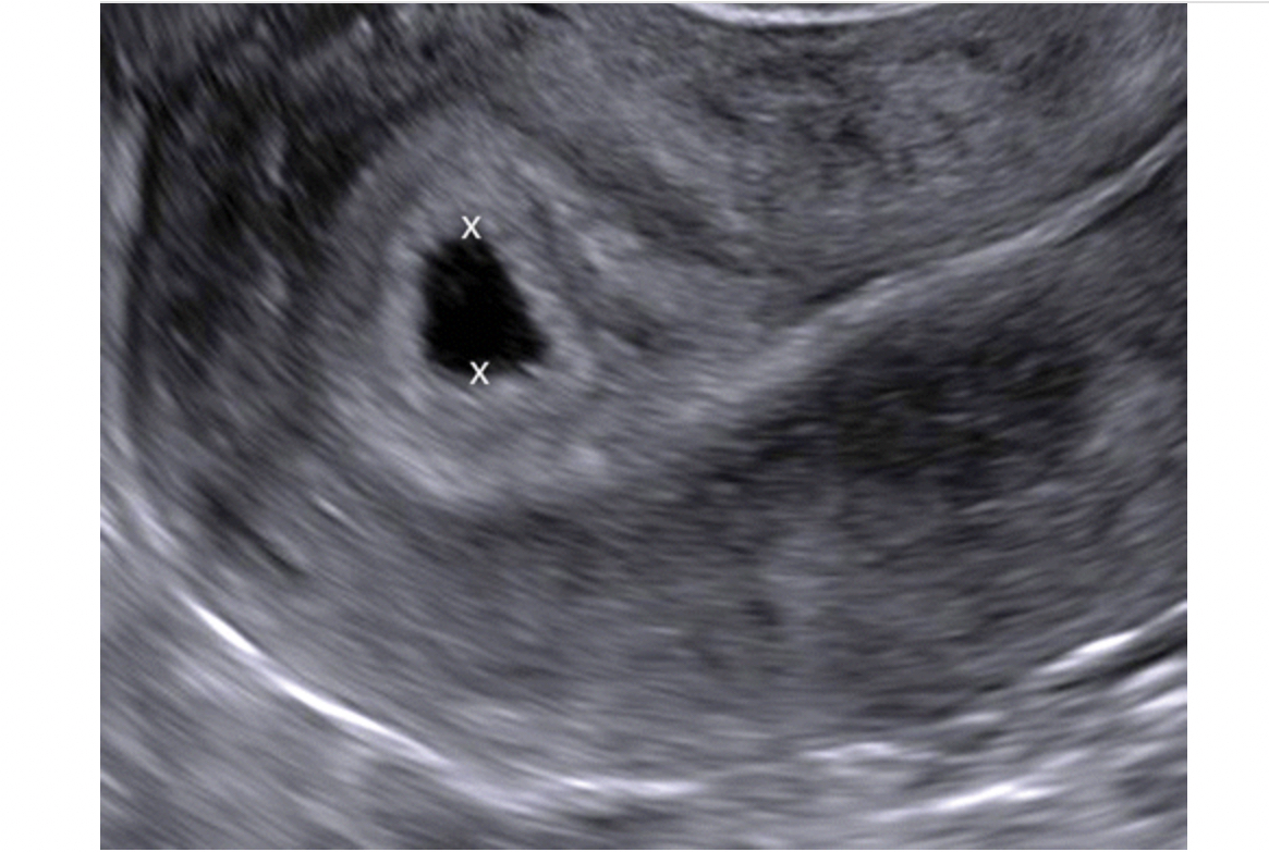
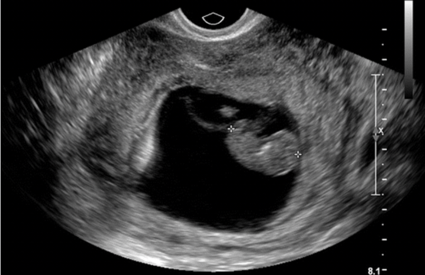
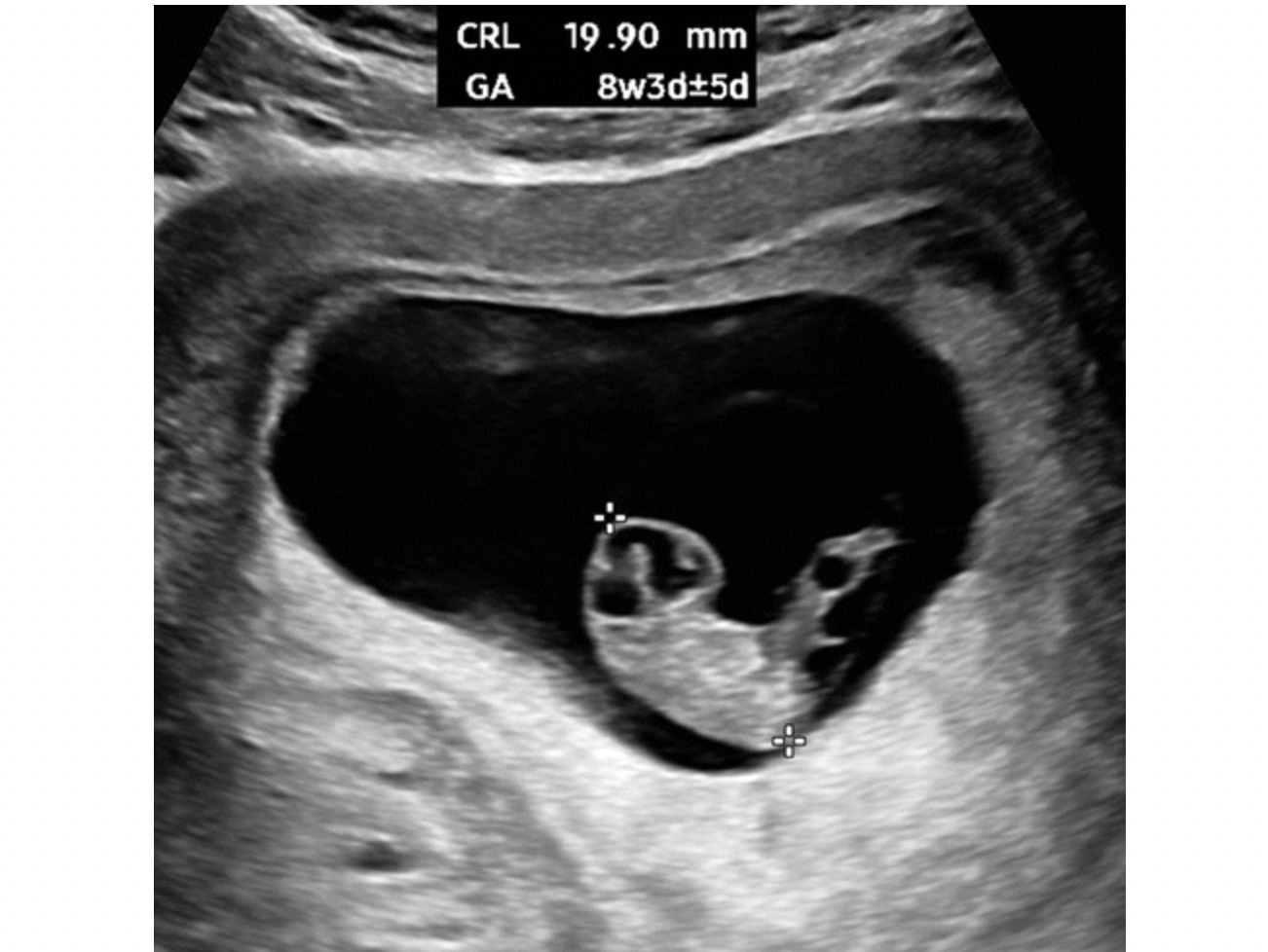
Early first trimester ultrasound measurements are used to calculate the gestational age of the pregnancy. This is the average of orthogonal diameters of the gestational sac, measured from inner border to inner border. An intrauterine sac–like structure without a yolk sac or embryo, as seen in this example, is described as a probable intrauterine pregnancy. Follow-up ultrasound is performed at 14 days, at which time a live embryo should be visible. Images courtesy of RSNA
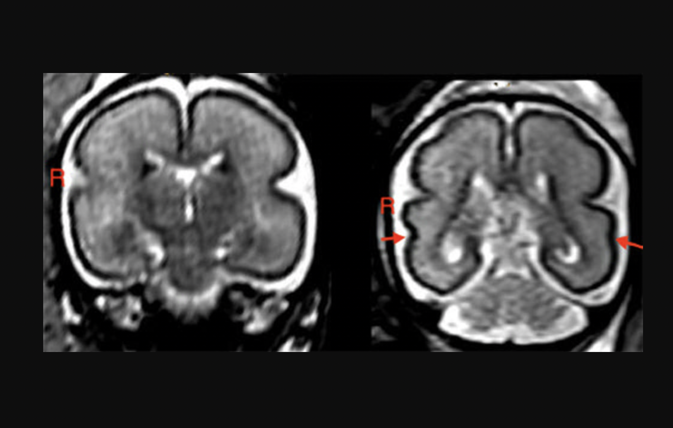
Alcohol consumption during pregnancy can change the unborn baby’s brain structure and delay brain development. Left: Fetal brain post-intrauterine alcohol exposure in fetus between 25 and 29 gestational weeks. Note the smooth cortex in the frontoparietal and temporal lobes. Right: Brain of matched healthy control case in fetus between 25 and 28 gestational weeks. The superior temporal sulcus is already bilaterally formed (red arrows) and appears deeper on the right hemisphere than on the left. Read more about this study.
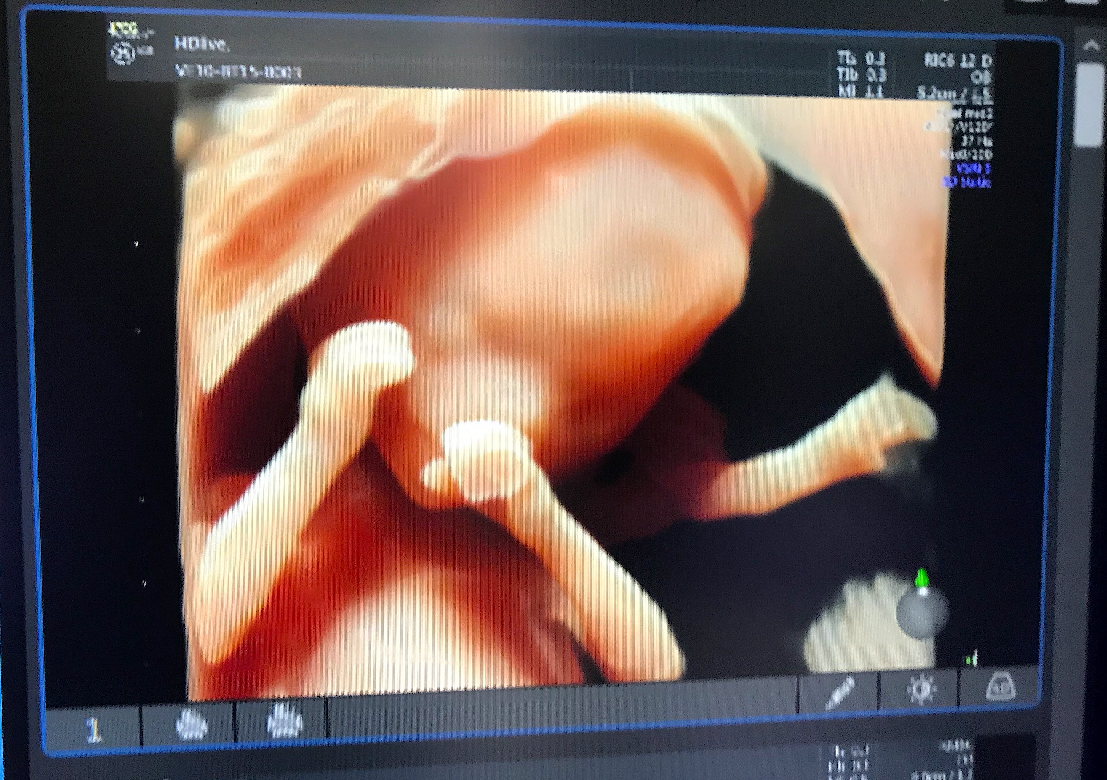
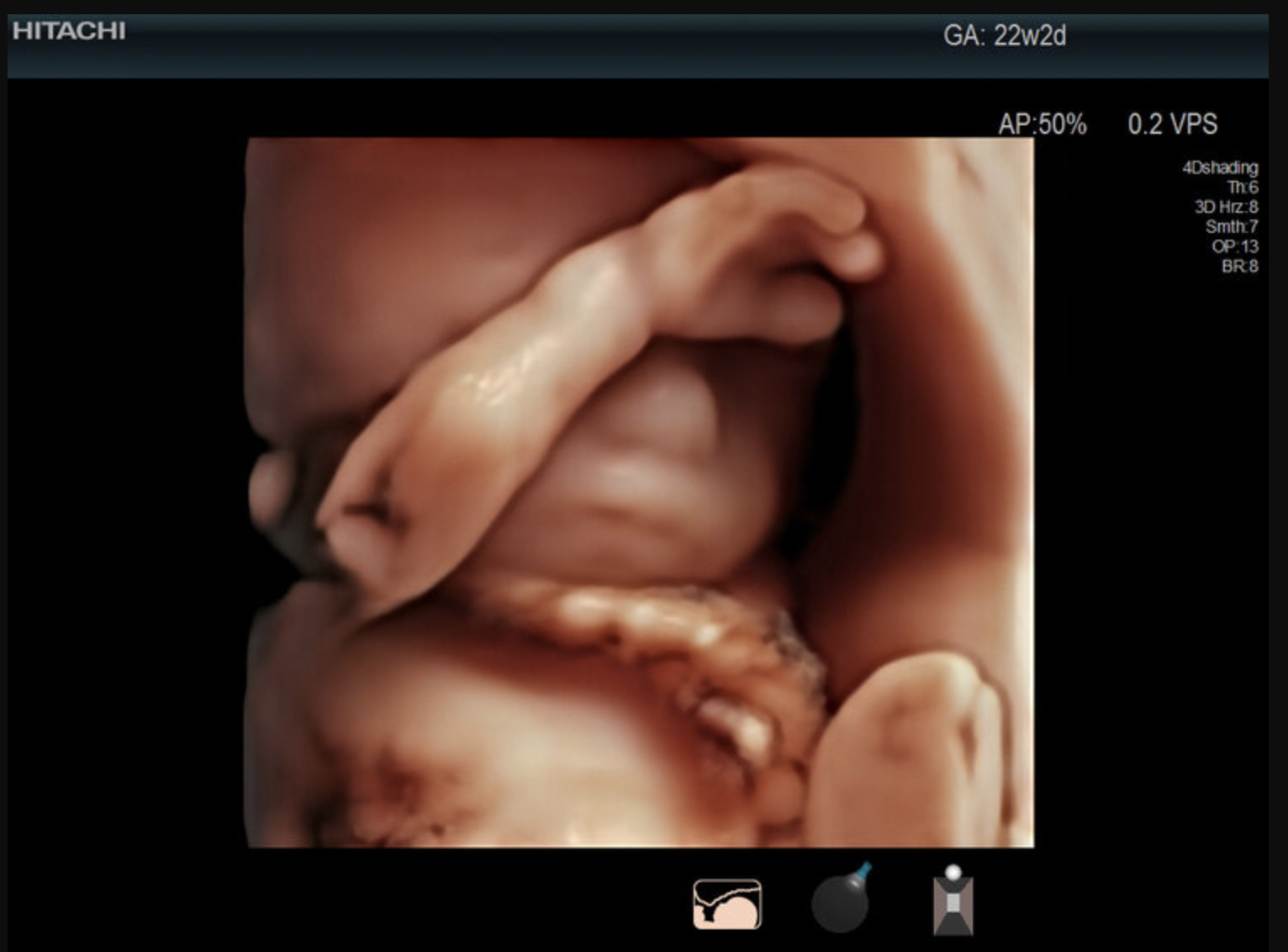
Fetal hand in a 3D ultrasound. Image courtesy of Hitachi.
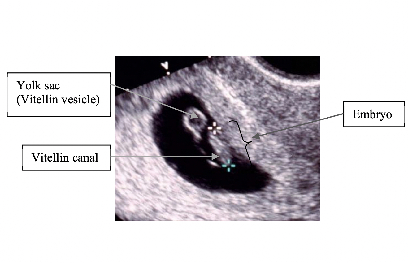
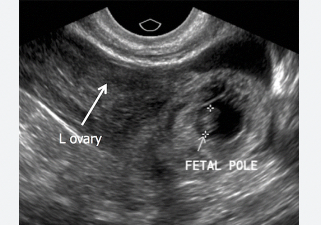
An example of a first trimester transvaginal ultrasound showing the sac and the embryo embedded on the edge of the uterine wall. Image courtesy of the Department of Global Health, University of Washington.
Third trimester fetal face and cerebral blood flow images with a Hitachi Prosound F37 ultrasound system. Ultrasound of pericallose artery in fetal brain with highly sensitive color mode (DFI). Image courtesy of Hitachi.
As a pregnancy advances, the embryo is easier to see, and accurate measurements can be obtained with transabdominal ultrasound. This ultrasound shows a crown-rump length (CRL) measurement with calipers. CRL is the average of discrete fetal measurements from the tip of the head end to the tip of the rump end in the midsagittal plane of the embryo. This is highly accurate for pregnancy dating in the first trimester. Images courtesy of RSNA.
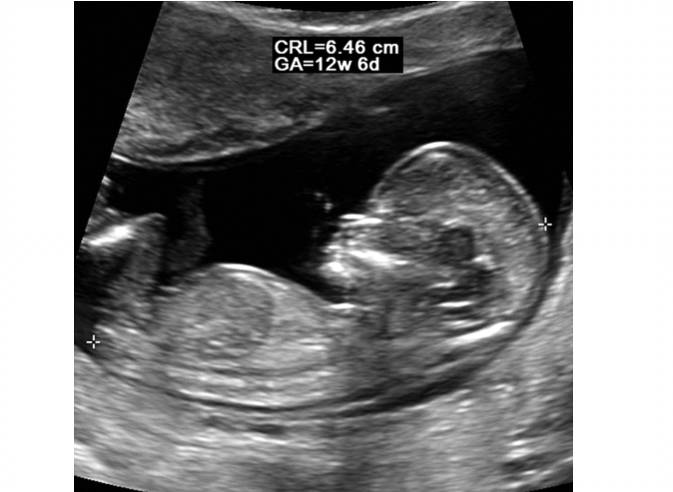
Ultrasound scan shows the fetus at the end of the first trimester, by which time the fetus has a recognizable human appearance. The maximal length in the craniocaudal direction is measured in a straight line between the calipers to make a crown-rump length (CRL) measurement. This is highly accurate for pregnancy dating in the first trimester, but other measures are used to assess gentational age later during the pregnancy. Images courtesy of RSNA.
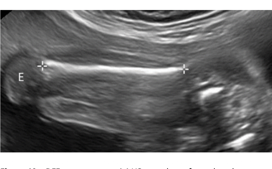
Ultrasound of a femur length measurement in a fetus at 24 weeks gestation. It shows the the epiphysis (E), the end part of the bone that initially grows separately from the shaft, without internal calcification. Bones in the fetus and babies ossifies into solid bone over time, allowing the bones to grow more rapidly. Image courtesy of RSNA.
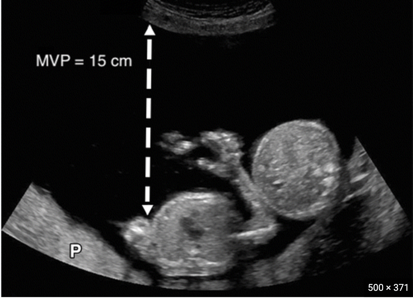
Ultrasound measurement of kidney length in a 23 week-old fetus. Renal measurements are one of several fetal anatomic structures that demonstrate consistent growth throughout the latter half of gestation, and measurements of these structures reliably correlate with gestational age (GA). This can be important in the second trimester and beyond as a more accurate measure of GA than body length. Image courtesy of RSNA.
Measurement of the fetal head to determine biparietal diameter (BPD), the cross-sectional diameter of the skull, which is also known as the fetal head circumference. Using a BPD index, it is possible to know the growth, weight and size of the fetus before birth. This needs to be measured between 13th and 20th week of pregnancy, because from the 20th week onwards, the index gradually loses its accuracy. Image courtesy of the Department of Global Health, University of Washington.
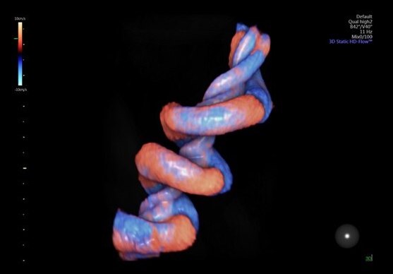
Very detailed view of the umbilical cord in a 3D/4D fetal ultrasound using the GE Healthcare HDlive Flow technology. The color code of the Doppler ultrasound shows blood flowing toward and away from the transducer to enable blood flow assessments.
Twin pregnancy, where the head of the second fetus can be seen on the right side and the rest of its body outside the imaging plane. Image courtesy of the Department of Global Health, University of Washington.
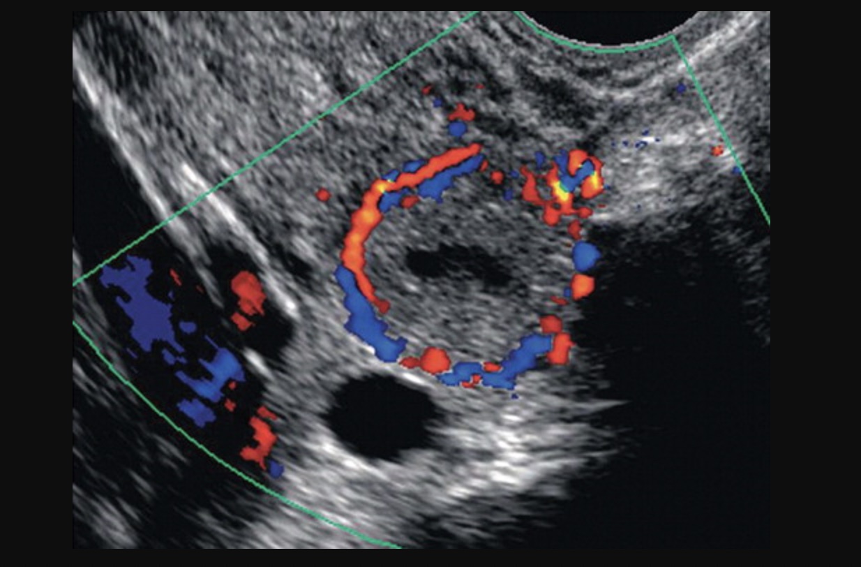
Ectopic pregnancy confirmed by Doppler ultrasound that shows a "ring of fire" sign caused by increased blood flow in a region. Ectopic pregnancy occurs when an embryo begins to develop outside of the uterus and can be dangerous for the mother. Ectopic pregnancies can occur in the fallopian tubes, ovaries and in the peritoneal space of the abdomen. Image courtesy of RSNA.
Ectopic pregnancy in a fallopian tube where the developing embryo can be seen inside the tube on ultrasound. Ectopic pregnancy occurs when an embryo begins to develop outside of the uterus and can be dangerous for the mother. Ectopic pregnancies can occur in the fallopian tubes, ovaries and in the peritoneal space of the abdomen. Image courtesy of RSNA.
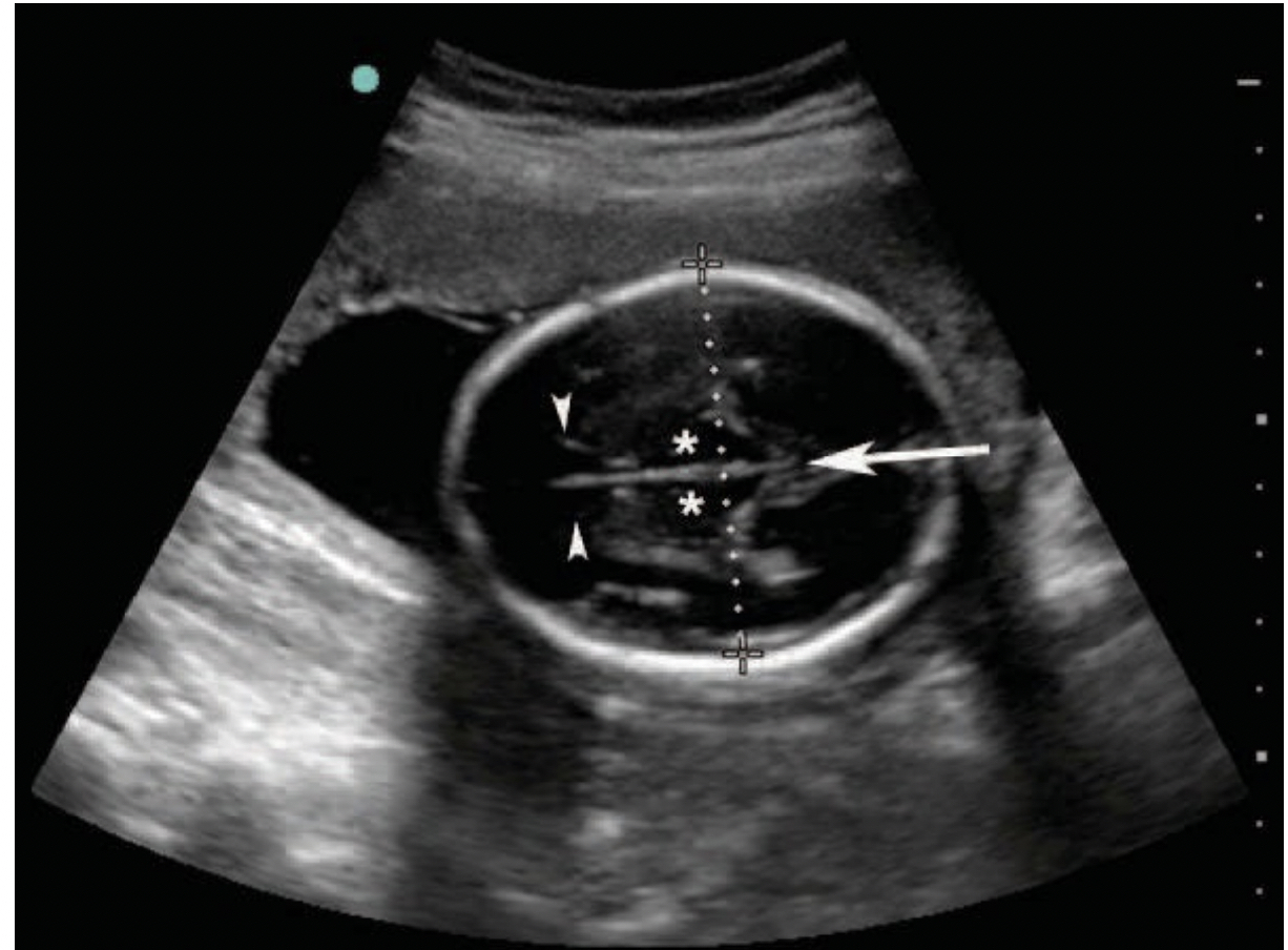
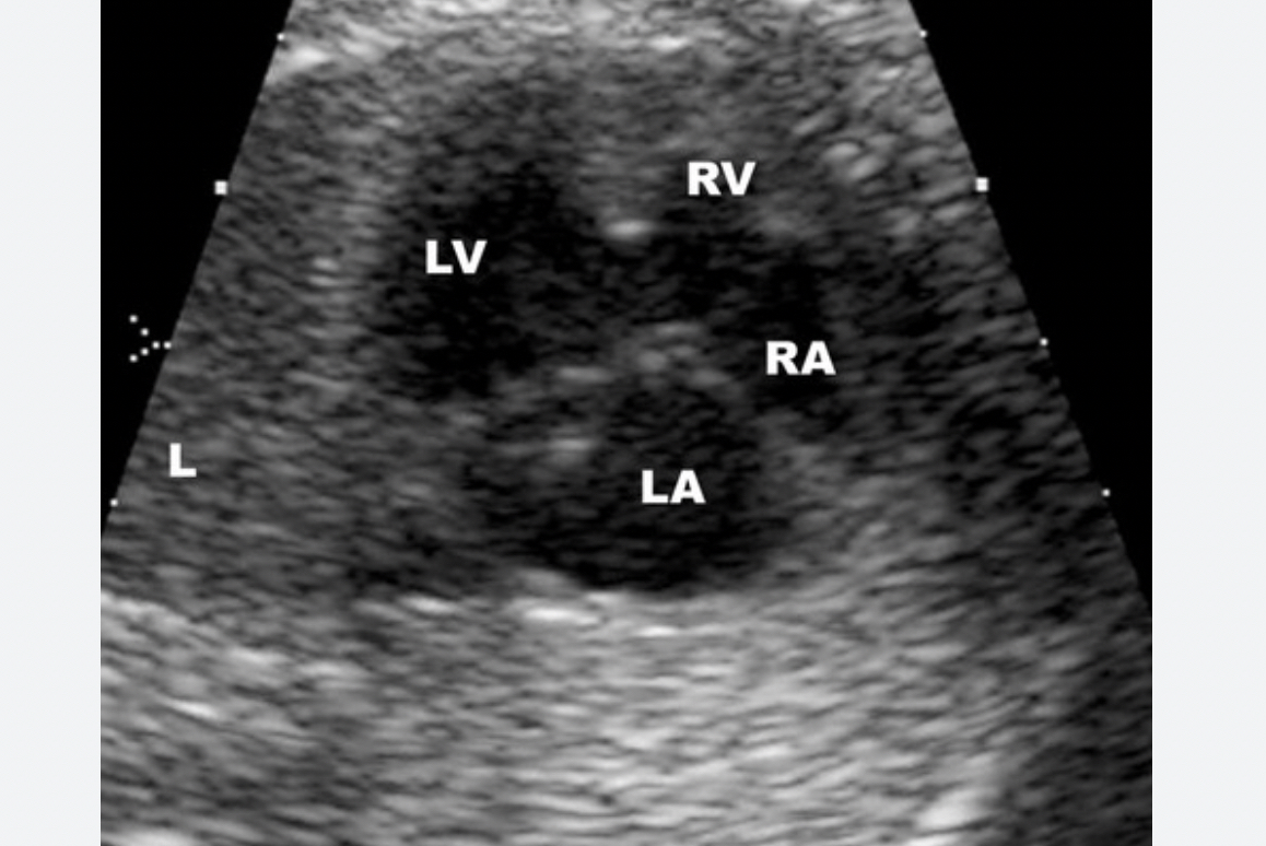
A standard four-chamber view of a fetal heart on ultrasound. The heart is examined to look for congenital heart disease (CHD), such as transposed vessels, connections between the chambers and defective heart valves. Image courtesy of RSNA.
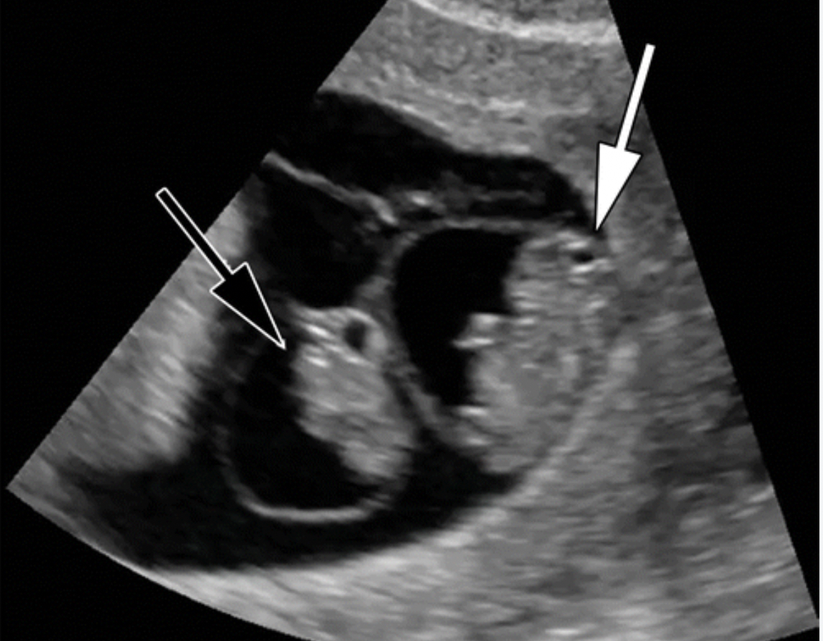
A congenital heart defect seen on a fetal ultrasound. The connection between the right and left ventricles has a ventricular septal defect (VSD), which can cause unoxygenated blood to be recirculated through the body. Image courtesy of RSNA.
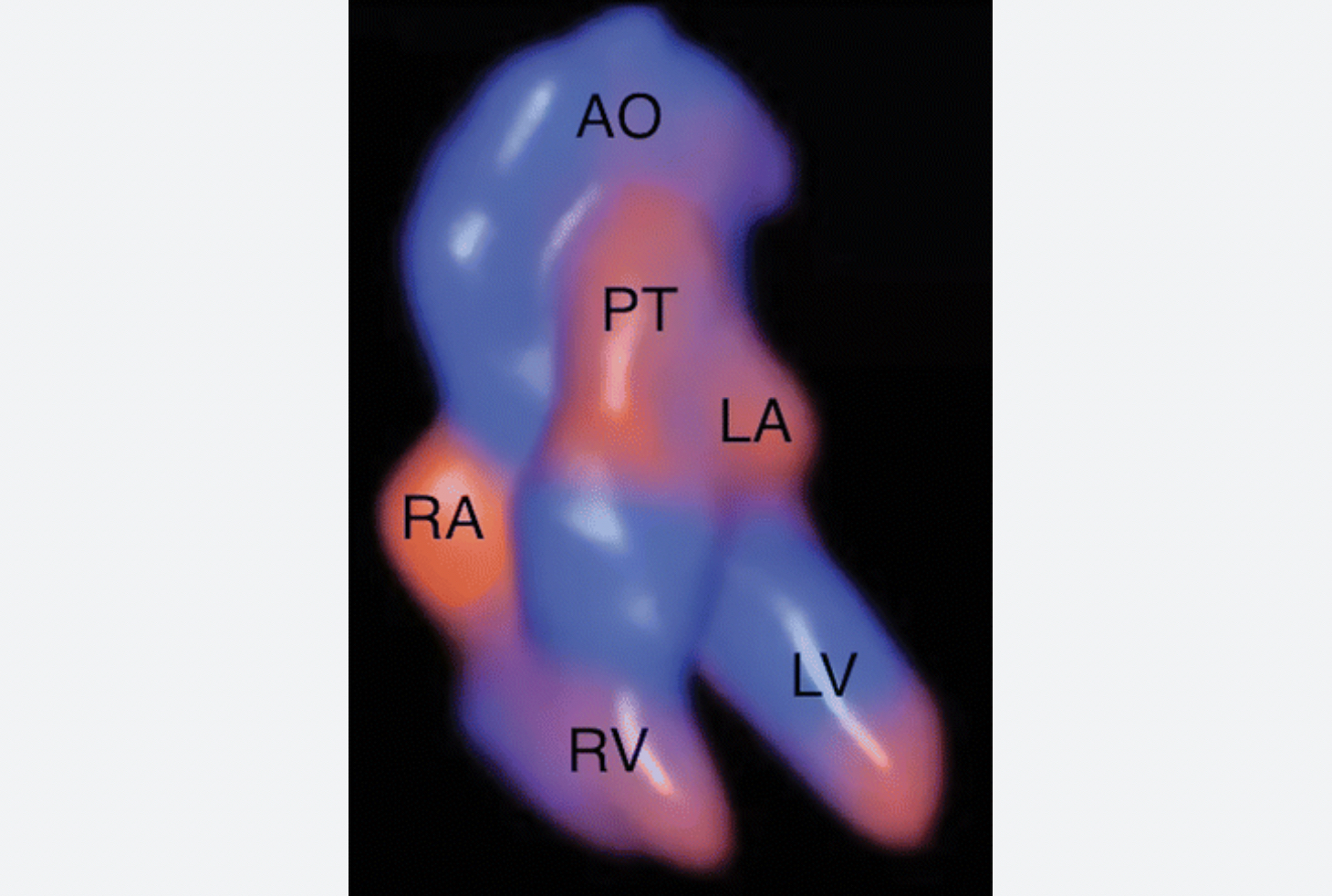
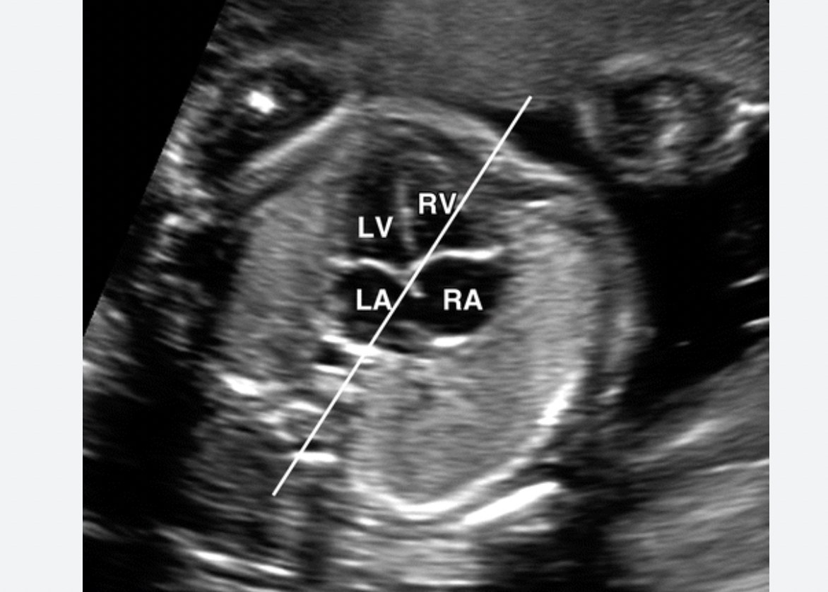
Four-dimensional “Glassbody” rendered image obtained in the Doppler cardiac spatio-temporal image correlation mode, showing the fetal cardiac anatomy at 16 weeks gestation. Retrograde flow is in red, and antegrade flow is in blue. This type of view may help earlier diagnosis of congenital heart disease in fetal imaging. AO = aorta, LA = left atrium, LV = left ventricle, PT = pulmonary trunk, RA = right atrium, RV = right ventricle. Image courtesy of RSNA.
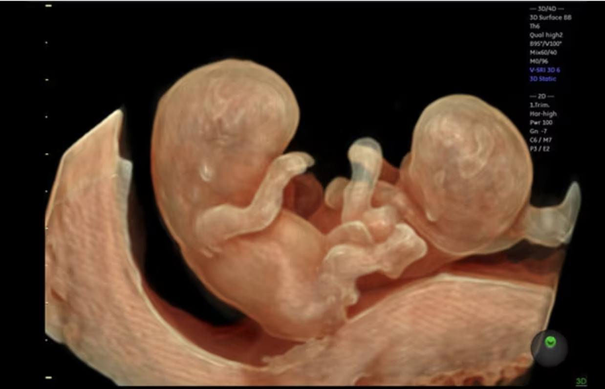
Twins seen in 3D fetal ultrasound. Image from GE Healthcare.
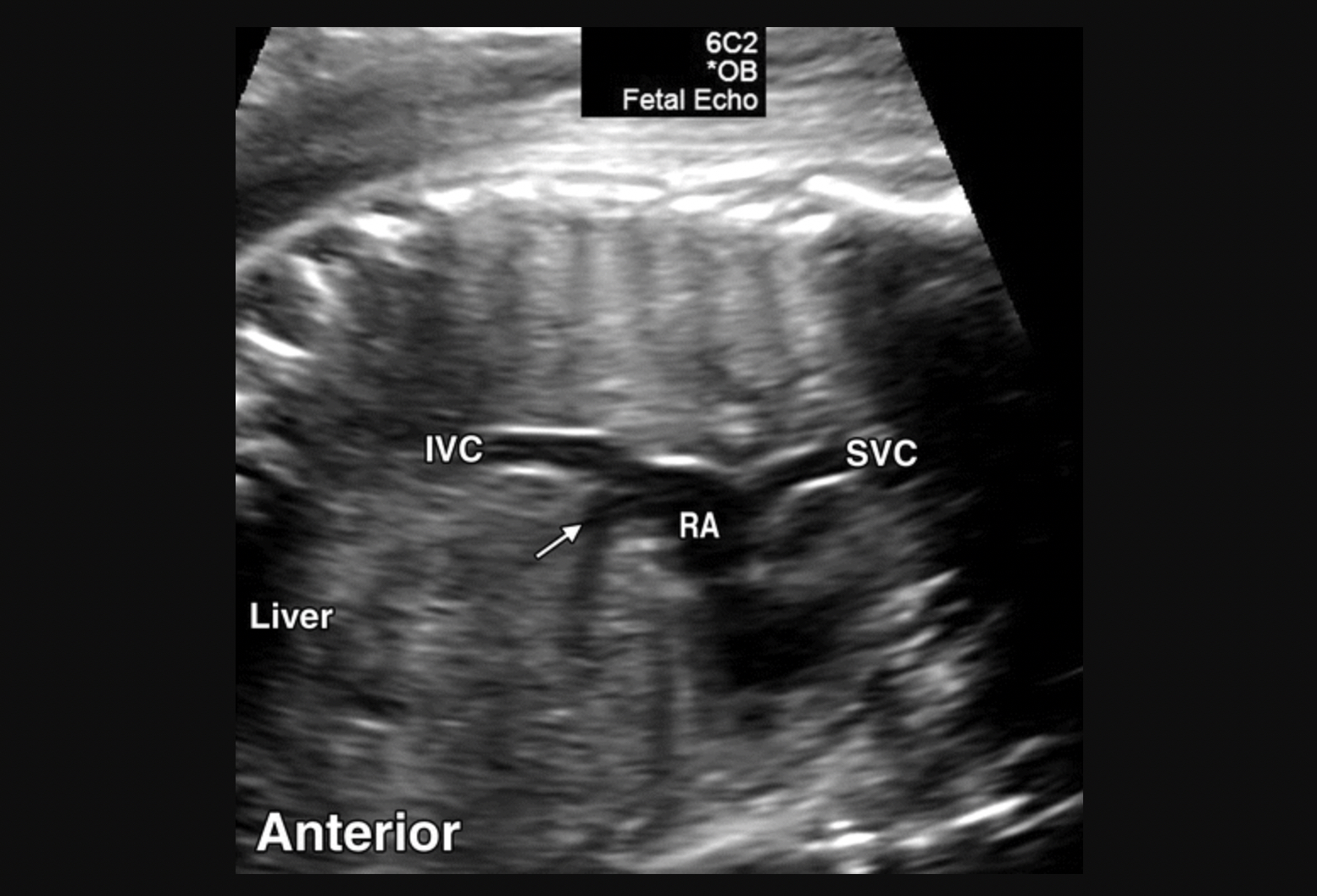
Fetal ultrasound evaluation of the vena cava connection into the right atrium of the heart to check for congenital heart abnormalities that merit referral for further evaluation. A similar evaluation will be performed on the aortic outflow tract of the heart to ensure there is normal anatomy. Image courtesy of RSNA.
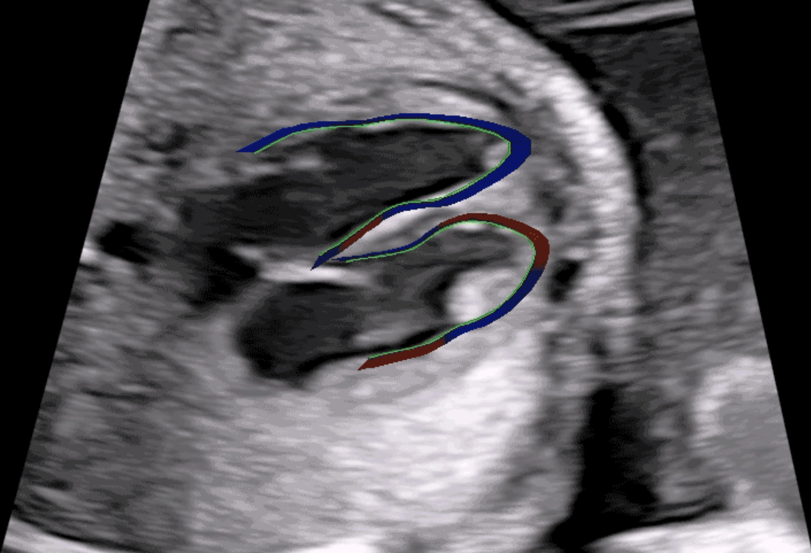
Example of a fetal heart contractility assessment ultrasound on the GE Voluson system. It divides fetal ventricles into 24 segments to simultaneously examine the size, shape and contractility of the fetal heart to help diagnose heart issues and congenital heart disease earlier in fetal development. Congenital heart defects affect 1 in 110 babies born around the world. Image from GE Healthcare.
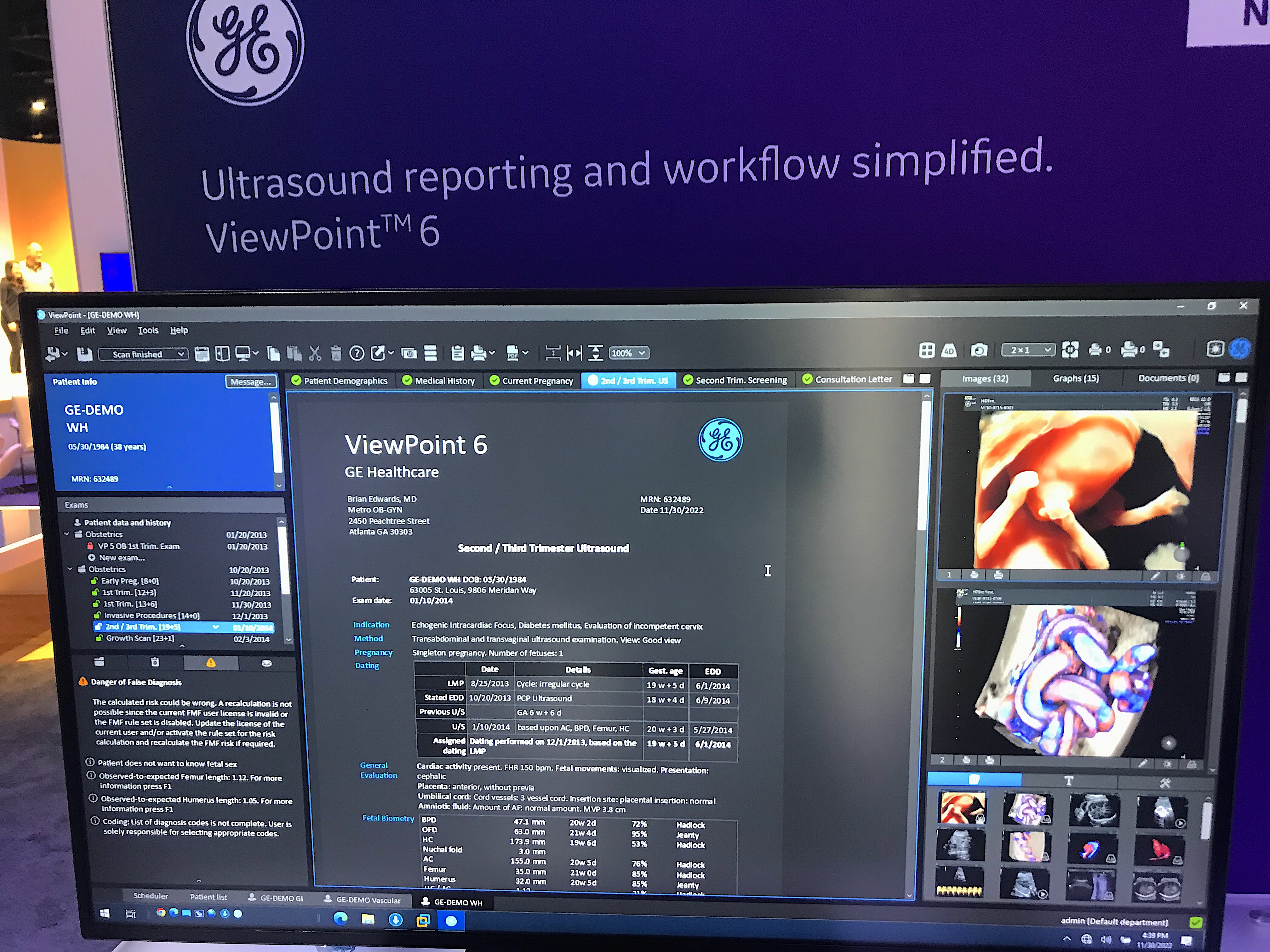
Example of a fetal ultrasound radiology report and the screen the physician sees. It shows the final report, notes in the patient's chart and images from the exam. Image from the GE Healthcare booth at RSNA 2022.
Example of a cleft lip detected deformity on 3D fetal ultrasound. Image courtesy of RSNA.
Twins on fetal ultrasound. Visible are the two heads and one of them has their arm outstretched. Image courtesy of RSNA.
Twins on fetal ultrasound in first trimester. Visible are the separate amniotic sacs, each with an embryo inside. Image courtesy of RSNA.
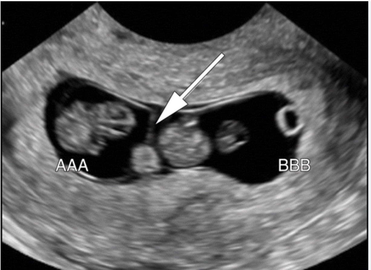
Twins on fetal ultrasound. The arrow points to the line showing the separate of the amniotic sacs. Image courtesy of RSNA.
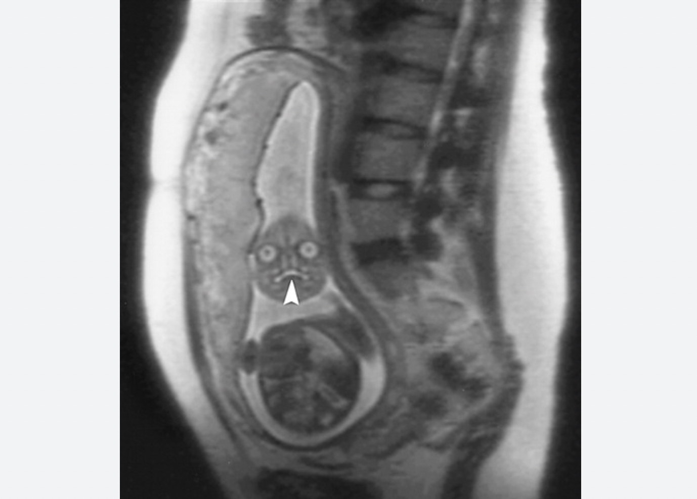
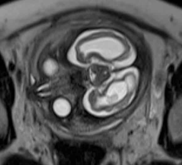
An MRI of a fetus inside the mother. The freakish appearance of the eyes and the face are normal for MRI fetal exams. Image courtesy of RSNA.
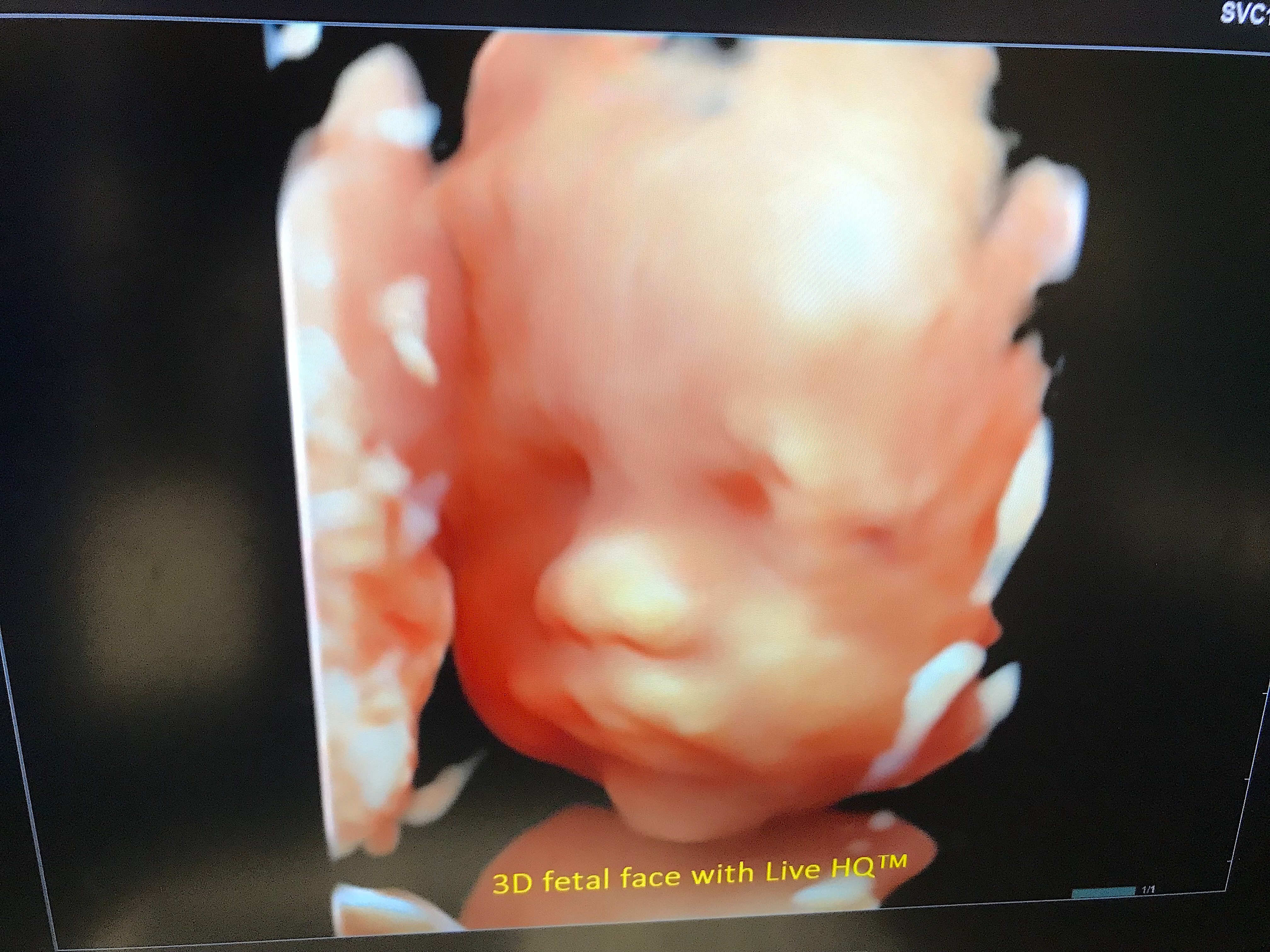
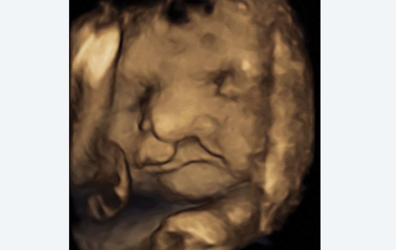
High definition 3D ultrasound of baby face. Image courtesy of Hitachi.
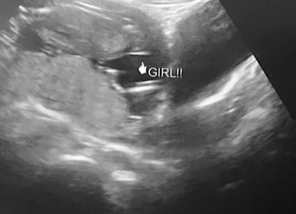
As a fetus develops, sonographers can identify the sex on ultrasound. This is an an example of a female and the "hamburger" sign of the vagina, which looks like the two buns of a hamburger or an equal sign. Males are identified by the "turtle" sign, where the genitalia look like a small turtle, or a turtle's head poking out.
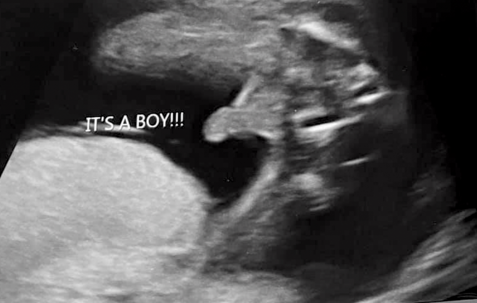
As a fetus develops, sonographers can identify the sex on ultrasound. This is an an example of the "turtle" sign, where the genitalia looks like a small turtle, or a turtle's head poking out. Females can be identifed by the "hamburger" sign of the vagina, which looks like the two buns of a hamburger or an equal sign.
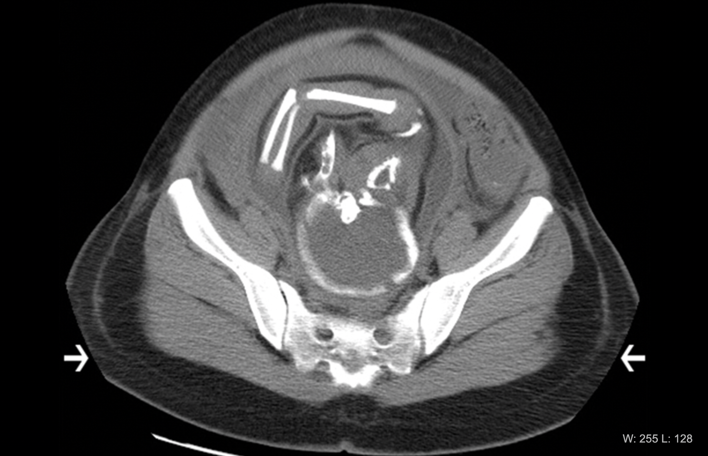
Sagittal computed tomography (CT) of a fetus inside the mother. CT is generally not used on pregnant women unless deemed necessary or in emergencies because of concerns about ionizing radiation dose to the developing fetus. For this reason, CT scans of fetuses are not common and there are usually protocols in place concerning when to image pregnant women with CT. Image courtesy of RSNA.
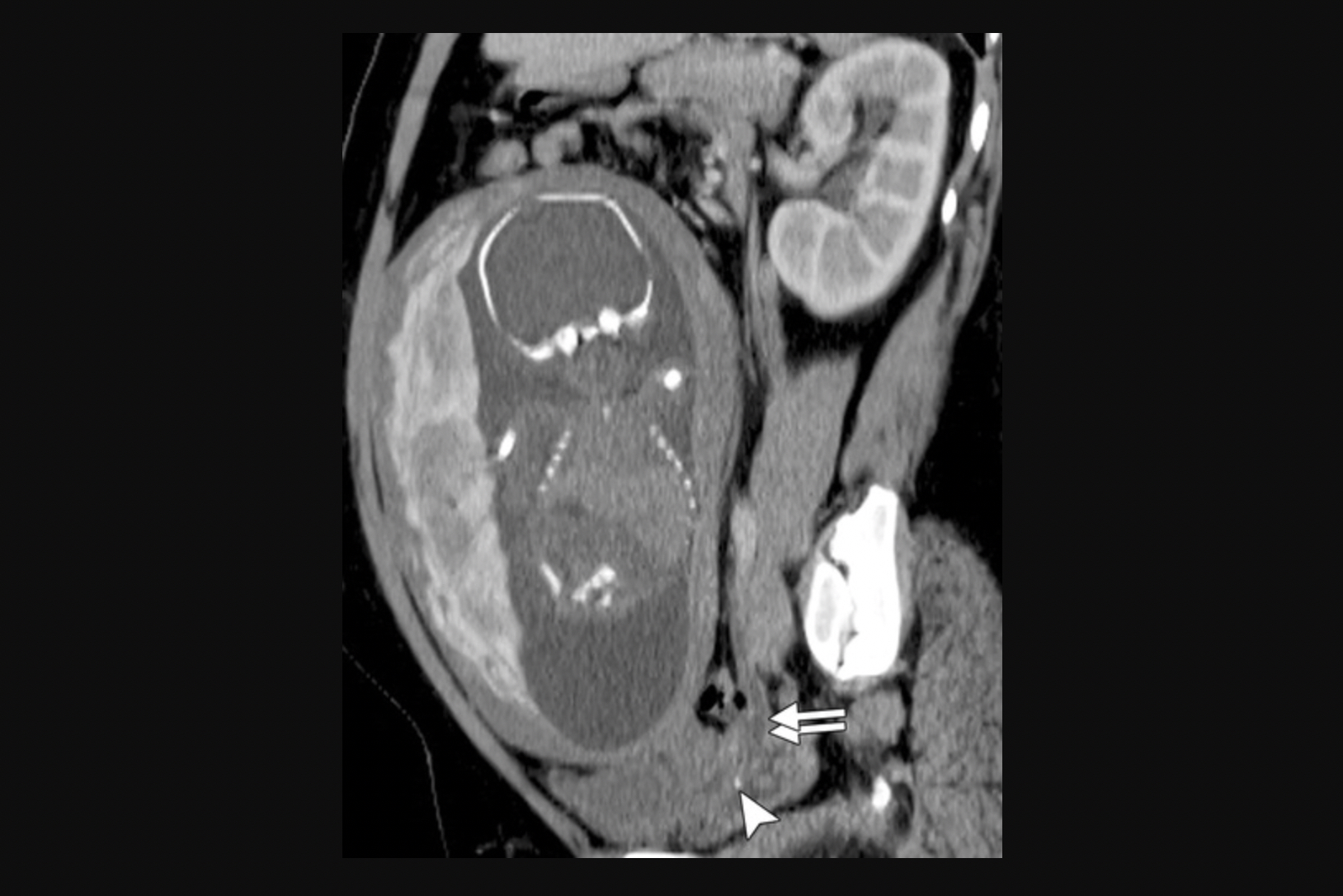
Axial computed tomography (CT) image of a third-trimester fetus. The patient was referred for CT by the emergency department because of right lower quadrant abdominal pain. Later-term pregnant patients often exceed the display field of view (arrows). The image shows a portion of the mother's hips and bottom of the spine (sacrum) and part of the baby's head and arm. CT is generally not used unless necessary on pregnant women because of concerns about ionizing radiation dose to the developing fetus. Image courtesy of RSNA.
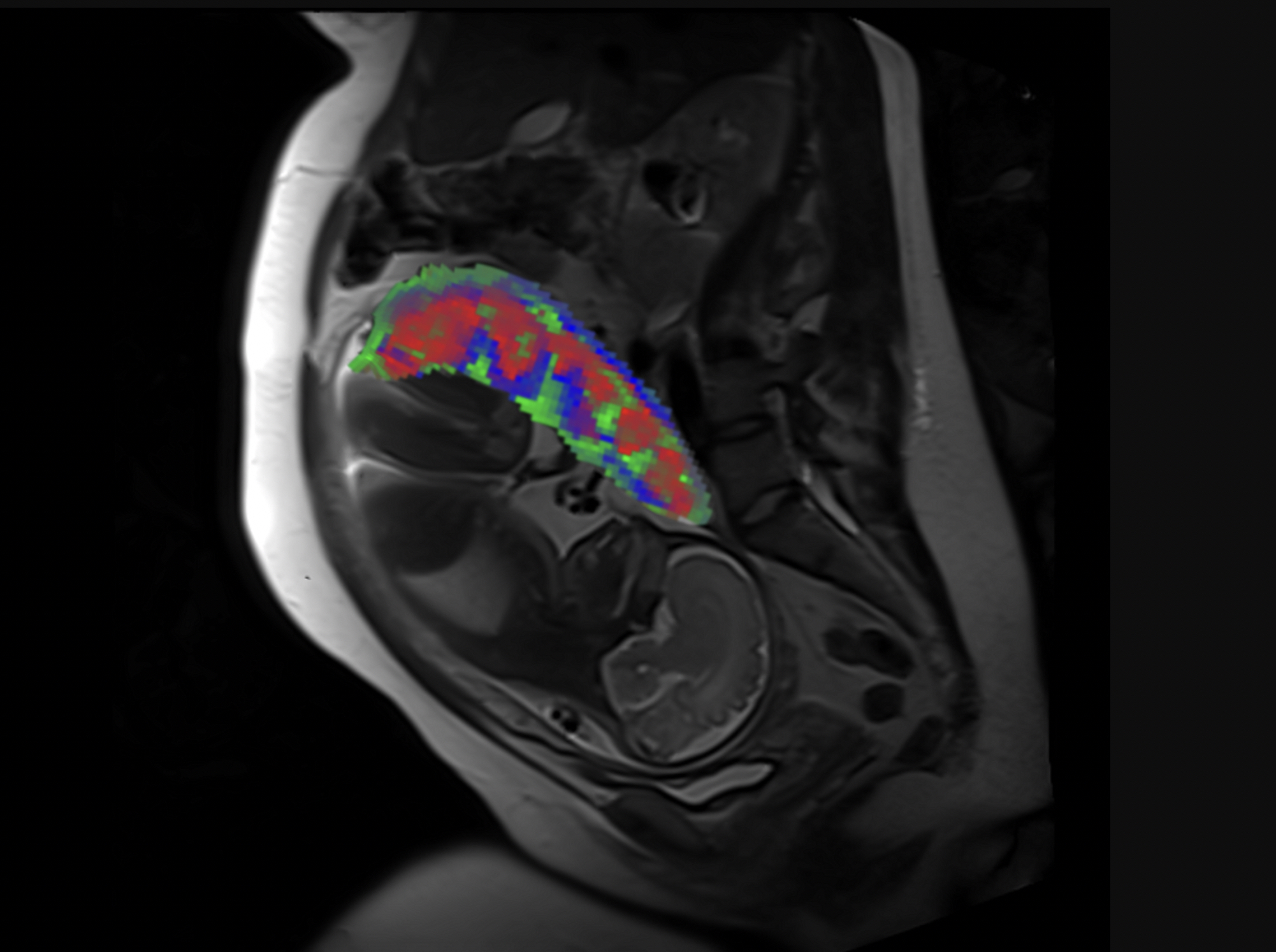
MRI scan showing the fetus and placental compartments—intervillous space (red), placental vessels (green), and placental tissue (blue). Researchers funded by the National Institutes of Health have developed a new method to process MRI scans to reveal the distinct compartments of the placenta, take measurements of oxygen levels in each region and determine if there are malformations in blood vessels (i.e., placental lesions). Obtaining this level of detail is currently not possible using standard MRI analysis methods. Image courtesy Wang Lab, Washington University in St. Louis
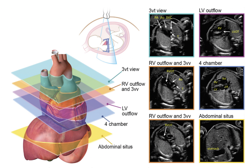
Axial imaging planes are suggested in the 2023 fetal echo guidelines for screening the fetal heart at the time of the obstetric anatomic survey and as an initial series obtained during fetal echocardiography. Read more
3D fetal ultrasound image of a 4-week old fetus images with a GE Voluson Expert 22 system, displayed at RSNA 2023.
Same image as the previous, but with an artificial lighting source that can be moved around the 3D ultrasound this 4-week old fetus images to give a life-like view, using an GE Voluson Expert 22 system. Rotation of the lighting source allows changes in contrast to help physicians better interpret the images.
Fetal umbilical cord hernia. Prenatal diagnosis is important to prevent inadvertent injury to the bowel during cord clamping at the time of delivery. This type of hernia can include a bulging of the intestines outside of the abdomen, but it is covered by the umbilical cord so may not be seen during delivery. This is a rare but important finding on prenatal ultrasounds, so a safe distance to clamp can be determined from the imaging and palpation of the cord during the time of delivery. Imaged using a GE Voluson Expert 22 ultrasound system.
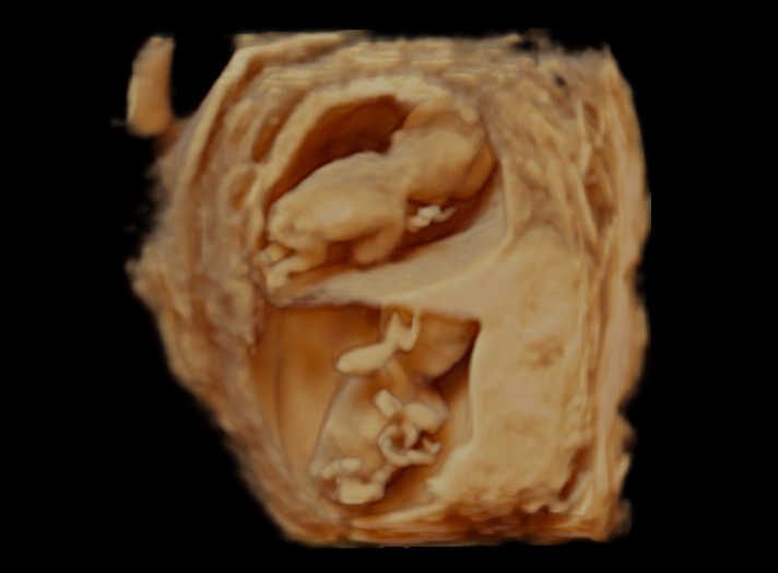
3D ultrasound images of first trimester fetal twins, scanned using a Canon Aplio 500.
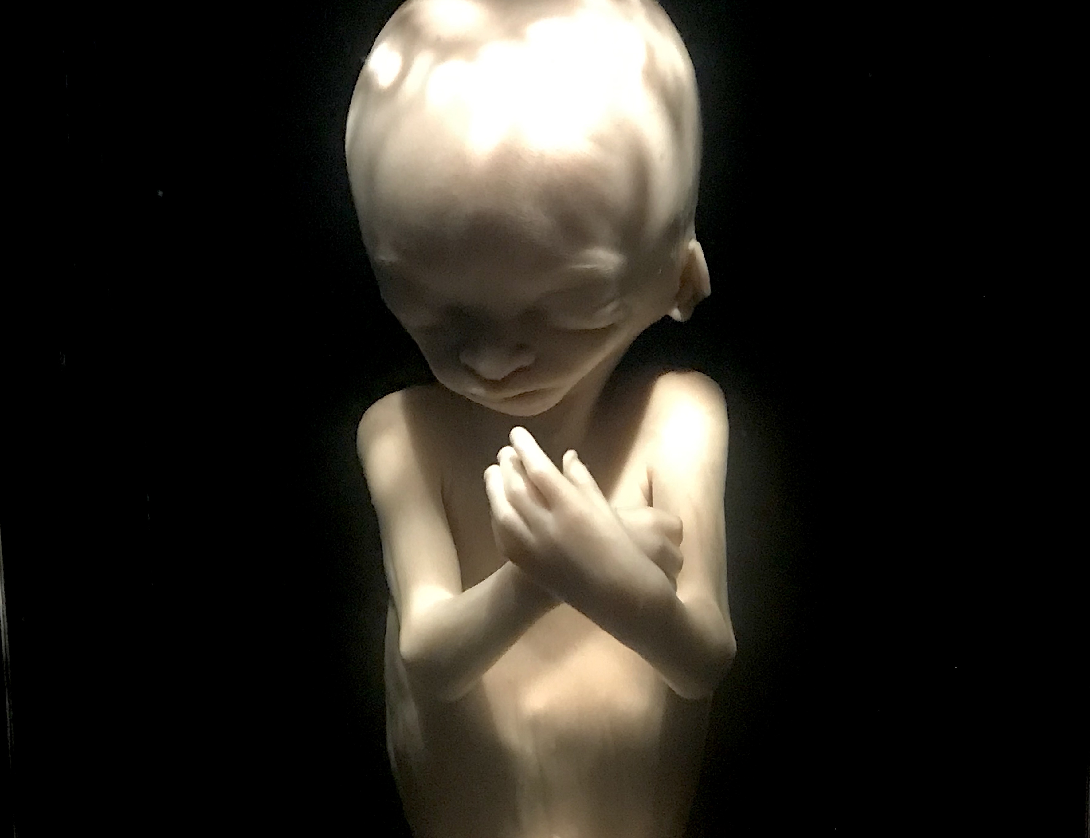
A preserved 14-week-old fetus inside the uterus on display as part of a permanent exhibit on the stages of fetal development at the Chicago Museum of Science and Industry. Link to museum's virtual tour. This image helps show correlation with fetal medical imaging. Photo by Dave Fornell.
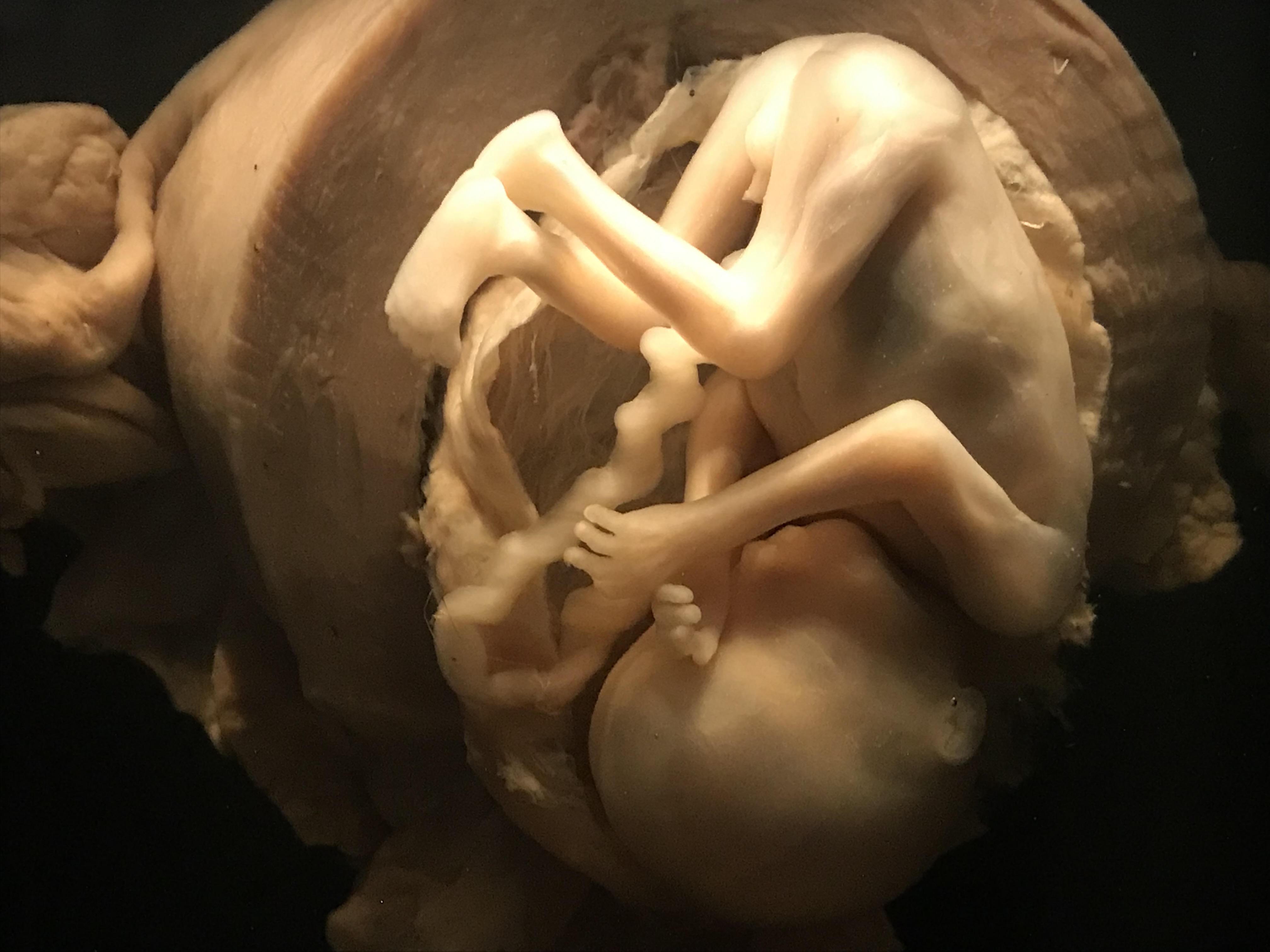
A preserved 18-week-old fetus on display as part of a permanent exhibit on the stages of fetal development at the Chicago Museum of Science and Industry. Link to virtual tour of the exhibit. This image helps show correlation with fetal medical imaging. Photo by Dave Fornell.
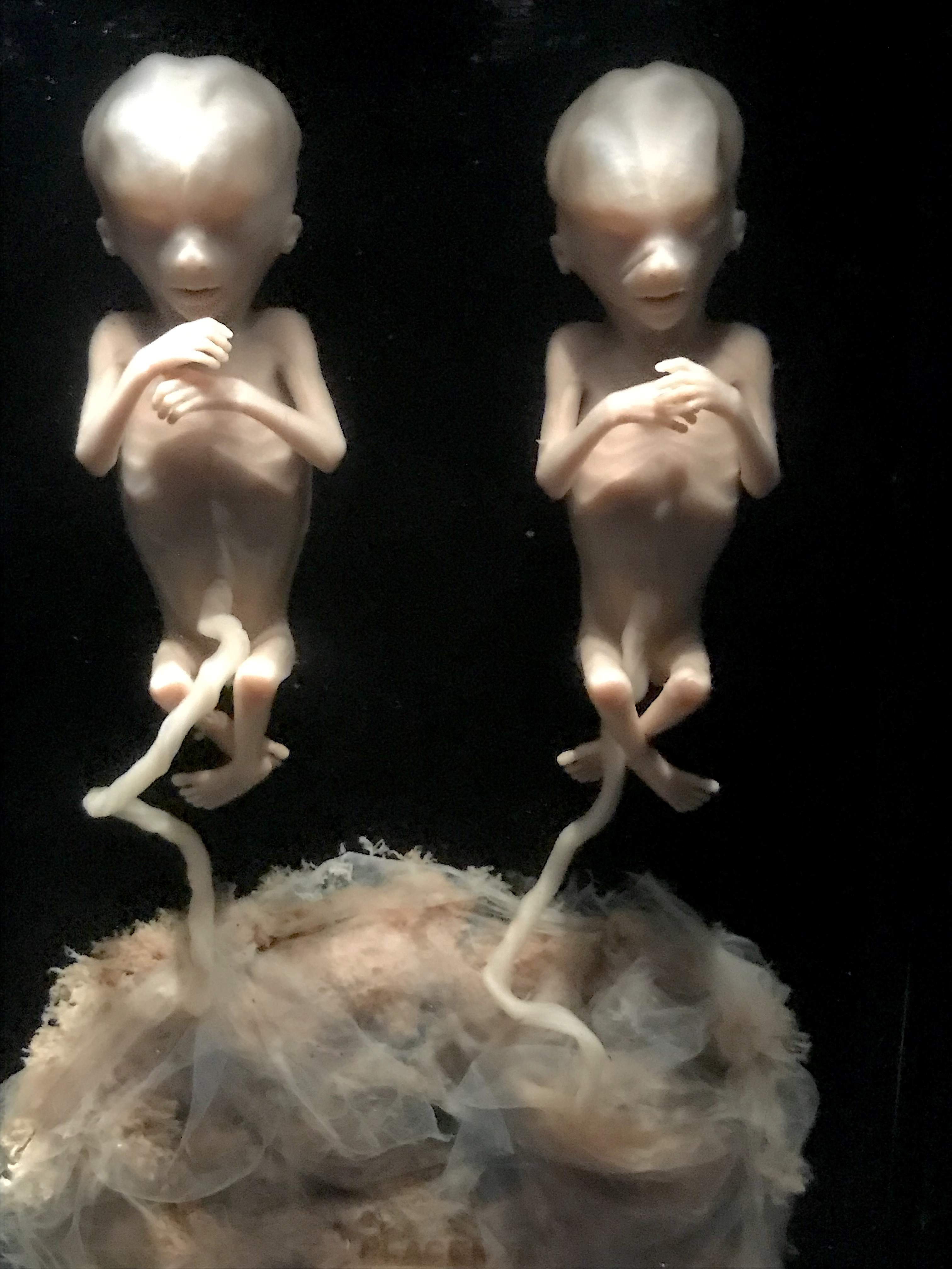
Preserved 13-week-old twin fetuses on display as part of a permanent exhibit on the stages of fetal development at the Chicago Museum of Science and Industry. These two fetuses developed when a single fertilized egg split, but both share the same genes and are identical twins. Link to virtual tour of the exhibit. This image helps show correlation with fetal medical imaging. Photo by Dave Fornell.

Dave Fornell has covered healthcare for more than 17 years, with a focus in cardiology and radiology. Fornell is a 5-time winner of a Jesse H. Neal Award, the most prestigious editorial honors in the field of specialized journalism. The wins included best technical content, best use of social media and best COVID-19 coverage. Fornell was also a three-time Neal finalist for best range of work by a single author. He produces more than 100 editorial videos each year, most of them interviews with key opinion leaders in medicine. He also writes technical articles, covers key trends, conducts video hospital site visits, and is very involved with social media. E-mail: dfornell@innovatehealthcare.com