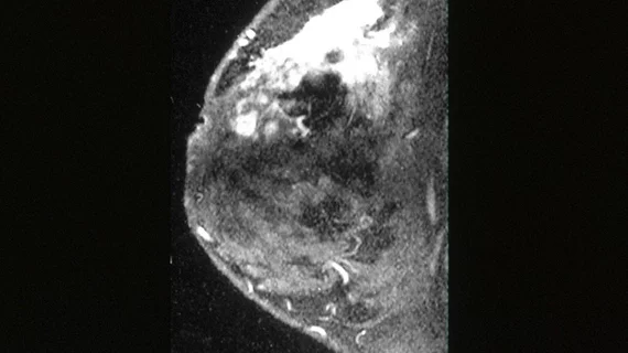
Which diffusion-MRI strategy best IDs malignant breast lesions?
Diffusion-weighted imaging techniques for quantitatively identifying benign and malignant breast lesions achieved comparable diagnostic accuracies, according to a meta-analysis of three methods published April 23 in Radiology.
“Whereas previous meta-analyses have assessed the performance of the (apparent diffusion coefficient) ADC model in differentiating between benign and malignant lesions, more advanced diffusion techniques aim to improve on the results of quantitative (diffusion-weighted imaging) DWI,” wrote Gabrielle C. Baxter from the department of radiology at the University of Cambridge School of Clinical Medicine in Cambridge, England, and colleagues.
The researchers looked at 73 eligible studies consisting of 3,930 malignant and 2,861 benign lesions that evaluated the diagnostic performance of DWI, diffusion-tensor imaging (DTI) and intravoxel incoherent motion (IVIM) in the breast.
These advanced diffusion techniques require “higher computational complexity and longer imaging time,” the authors wrote. If they were more accurate, such a shortcoming would be justifiable.
Overall, DWI, DTI and IVIM achieved comparable diagnostic performances for identifying breast malignancy at MRI, the authors wrote. DTI achieved a sensitivity, specificity and area under the curve of 93%, 90% and 0.94, respectively. IVIM was slightly below, scoring an 88% sensitivity, 79% specificity and AUC of 0.90 for identifying malignant versus benign lesions.
Diffusion-weighted MRI achieved an 89% sensitivity and 82% specificity and area under the curve of 0.92 for depicting malignant lesions, comparable to dynamic contrast agent-enhanced MRI, the authors noted.
“The pooled estimates of sensitivity, specificity, and area under the receiver operating characteristic curve were found to be comparable for ADC (commonly calculated using DWI), IVIM, and DTI,” Baxter and colleagues concluded. “However, because of the small number of studies included and the large confidence intervals, this meta-analysis lacks the statistical power to conclude that they are diagnostically equivalent.”

