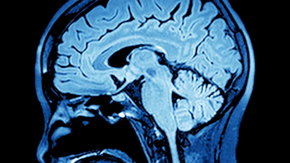
Magnetic resonance elastography advances epilepsy diagnoses
Using magnetic resonance elastography can help clinicians detect and diagnose epilepsy, researchers reported recently in NeuroImage: Clinical.
The findings are the product of a collaboration between the Carle Neuroscience Institute and the Beckman Institute for Advanced Science and Technology at the University of Illinois, both located in Urbana-Champaign.
Current approaches to spotting mesial temporal lobe epilepsy—the most common form of the disease that’s resistant to treatment—usually involve MRI and often fall short. MRE, on the other hand, can detect early changes that are critical to treating the condition.
"The structural changes in the brain, in response to seizures, cause the death of neurons and the formation of scar tissue," Graham Huesmann, a neurologist at Carle, said in a statement on Sept. 2. "By the time we see any changes on the MRI, the disease is pretty advanced. We wanted to detect these changes earlier using MRE."
The technique involves having an individual lie on a small vibrating pillow, monitoring the changes as they move through tissue, and encounter various compositions and organizational abnormalities, the authors explained. It’s already well-regarded for staging liver diseases and for completing liver biopsies.
For their research, Huesmann et al. used MRE to noninvasively analyze hippocampal stiffness in 12 individuals with unilateral MTLE and 13 healthy patients. After analysis, they noted a higher brain stiffness ratio in those with epilepsy compared to controls.
The group said adding MRE to neuroimaging assessments may dramatically help patients better understand their specific disease type. They’re now planning to refine the process to look at additional forms of epilepsy.
"All of our imaging techniques currently depend on looking at brain chemistry and the static images of the brain," Huesmann added. "Using MRE to see how the brain jiggles is an exciting way to approach this problem. It is also an inexpensive technique and can therefore be used by anyone."
Read the entire study here.

