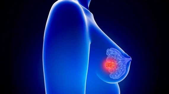
AI triages breast screening MRIs without missing a single cancer
A newly developed automated system can quickly assess MRI exams of women with dense breasts and toss those without cancer, ensuring radiologists can focus on more difficult cases.
Patients with dense breast tissue face a twofold higher risk of developing cancer than most women. Adding supplemental MRI to normal screening has proven benefits for this population, so researchers used this imaging data to develop a deep learning tool.
Putting it to the test on more than 9,000 dense breasts, AI triaged about 90% for radiologists’ review due to abnormal lesions. Nearly 40%, meanwhile, did not have lesions and were later confirmed cancer-free.
“We showed that it is possible to safely use artificial intelligence to dismiss breast screening MRIs without missing any malignant disease,” Erik Verburg, MSc, from the Image Sciences Institute at the University Medical Center Utrecht in the Netherlands, said Tuesday. “The results were better than expected. Forty percent is a good start. However, we have still 60% to improve.”
To create their tool, Verburg and co-investigators used 4,500 MRI datasets from a Dutch study underscoring the benefits of supplemental MRI screening for women with dense breasts. Overall, this included scans from seven hospitals and testing data from an eighth.
In addition to its 100% sensitivity for cancerous lesions, AI notched an average area under the receiver operating characteristic curve of 0.83.
More than half of women are willing to shell out at least $250-$500 for a screening MRI, particularly those with dense breasts, one recent study found. Having an automated system sort through these scans may significantly help busy rads, the Dutch authors noted.
“The approach can first be used to assist radiologists to reduce overall reading time,” Verburg added. “Consequently, more time could become available to focus on the really complex breast MRI examinations.”
Despite its clinical benefits, breast MRI faces plenty of hurdles, including issues surrounding workflow, reimbursement, quality and safety. And in the U.S., federally-certified physicians are required to interpret such exams, Bonnie N. Joe, a radiologist with the University of California, San Francisco noted.
You can read the entire study published in Radiology here, and the editorial from Bonnie N. Joe here.

