Experts spot structural differences on brain imaging of individuals with autism
Experts are inching closer to being able to objectively understand how the brains of individuals with autism spectrum disorder (ASD) function differently from their neurotypical peers.
New research from the University of Virginia suggests that the microstructures of the brains of those with autism differ from those not considered on the spectrum. This, experts involved in the study explained, offers further insight into exactly what is happening inside the brain that causes individuals on the spectrum to process information differently.
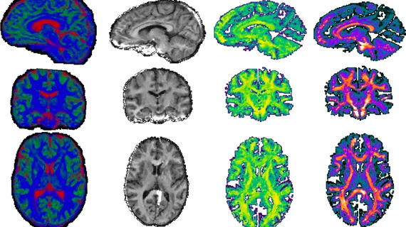
Scientists used a special type of MRI exam—diffusion tensor imaging—to analyze the water that continuously moves throughout the brain and cell membranes, while paying close attention to the myelin and axons. Through this, researchers were able to calculate individuals’ axonal conduction velocity, which offers insight into an axon’s ability to transfer information throughout the brain.
When comparing the imaging of people on the spectrum to others who are otherwise considered neurotypical, the team found significant differences in the extracellular water and conduction velocity throughout the cortex, subcortex and white matter skeleton between the two groups, with the ASD group showing increased water presence and decreased conduction velocity.
The group used these metrics to create mathematical models of the brains’ microstructures, which, in turn, allowed them to identify notable structural differences between the two groups. The participants’ scores on the Social Communication Questionnaire—a tool commonly used when diagnosing ASD—were found to be directly related to the microstructural differences observed on imaging.
“What we're seeing is that there's a difference in the diameter of the microstructural components in the brains of autistic people that can cause them to conduct electricity slower,” lead author of the paper, Benjamin Newman, a postdoctoral researcher with UVA’s Department of Psychology, said in a release. “It's the structure that constrains how the function of the brain works.”
Co-author John Darrell Van Horn, a professor of psychology and data science at UVA, highlighted the objective nature of the study, pointing out that ASD is typically diagnosed based on a pattern of behaviors, which can be quite subjective depending on who is conducting the examination.
“We need greater fidelity in terms of the physiological metrics that we have so that we can better understand where those behaviors come from,” Van Horn said. “This is the first time this kind of metric has been applied in a clinical population, and it sheds some interesting light on the origins of ASD.”
The authors suggested that this research could lay the foundation for identifying biological targets on which experts could focus future treatments and therapies, providing an objective means of measuring effectiveness.

