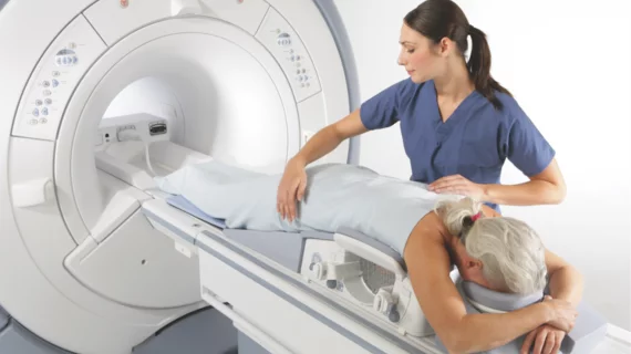
While promising, machine learning still misses 20% of cancers on breast MRIs, analysis shows
Although promising, the benefits of using machine learning-based MRI for detecting axillary lymph node metastases in breast cancer patients fell short of earning expert recommendation, according to a new study published in Insights into Imaging.
Diagnosing axillary lymph node metastases (ALNM) can be difficult, but its detection is crucial in a patient’s treatment plan and prognosis. In recent years, MRI has emerged as a valuable imaging modality in breast cancer research, and MRI in conjunction with machine learning (ML) models has been shown to detect ALNM. However, until now, a thorough analysis of its accuracy had not yet been conducted.
For this meta-analysis, experts pooled all studies from PubMed, Embase, Web of Science and Cochrane Library that examined the use of machine learning-based MRI models to diagnose ALNM in breast cancer patients. Fourteen articles were selected, totaling 2,247 patients with known breast cancer. Of those individuals, 1,508 had normal lymph nodes, while 954 had abnormal nodes.
Overall, the AUC for ML in the validation set was 0.80 and had a negative predictive value of 0.83. When analyzing the subgroup of the validation set, T1-weighted contrast-enhanced (T1CE) imaging with ML showed increased sensitivity compared to the T2-weighted fat-suppressed (T2-FS) imaging and diffusion-weighted imaging (DWI).
Although this research indicates that 80% of patients could avoid invasive biopsy or surgery to test lymph nodes, 20% of patients could receive a false negative result. Experts also pointed out that the performance of ML varied depending on MRI sequences and algorithms.
While machine learning-based MRI is a valuable and noninvasive tool for the detection of ALNM, it should not yet be considered in clinical routine, experts suggested.
“Although its results were encouraging with the pooled sensitivity of around 0.80, it meant that 1 in 5 women would go with undetected metastases, which may have a detrimental effect on the overall survival,” Chen Chen, with the Department of Radiology at West China Hospital, Sichuan University, and co-authors wrote.
You can read the detailed study in Insights into Imaging.

