
MRI study brings scientists closer to diagnosing CTE in living patients
Chronic traumatic encephalopathy cannot currently be diagnosed in living patients. But with the help of MRI researchers are now one step closer to achieving this goal, according to a study recently published in Alzheimer's Research and Therapy.
A neurodegenerative tauopathy, chronic traumatic encephalopathy (CTE) is a result of repetitive impacts to the head. It is commonly found in athletes who play contact sports, military personnel who have been in combat and abuse victims postmortem.
Due to a lack of validated in vivo biomarkers that can differentiate CTE from other neurological pathologies, the diagnosis cannot yet be confirmed in living patients. The authors cite the progress of previous studies that involve positron emission imaging and cerebrospinal fluid protein analysis but stress the degree of complexity involved.
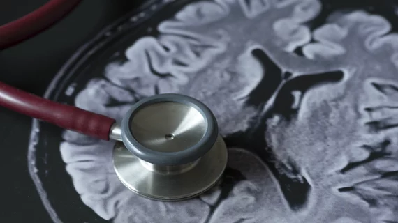
MRI is a valuable diagnostic tool in these instances, as it is common, cost-effective and readily available. With the help of magnetic resonance images conducted while patients with suspected CTE are still living, comparisons between antemortem and postmortem brains with autopsy-confirmed CTE can be made.
“MRI is commonly used to diagnose progressive brain diseases that are similar to CTE, such as Alzheimer’s disease. Findings from this study show us what we can expect to see on MRI in CTE,” wrote Jesse Mez, MD, director of the Boston University Alzheimer’s Disease Center Clinical Core, and colleagues.
For this study, the MRIs of 55 men with confirmed and symptomatic CTE were analyzed alongside 31 men considered to have “normal cognition.” The donors with confirmed CTE exhibited greater atrophy of the orbital-frontal, dorsolateral frontal, superior frontal, anterior temporal and medial temporal lobes. These donors also were shown to have significantly higher visual MRI rating scores for the lateral ventricles and third ventricle.
“The current findings provide, for the first time, insight into the structural MRI profiles of people with neuropathologically confirmed CTE, as well as support p-tau accumulation as a correlate of atrophy in CTE,” explained Michael L. Alosco, PhD, with Boston University Alzheimer’s Disease Research Center and CTE Center, and co-authors.
You can read the detailed research here.

In addition to her background in journalism, Hannah also has patient-facing experience in clinical settings, having spent more than 12 years working as a registered rad tech. She began covering the medical imaging industry for Innovate Healthcare in 2021.