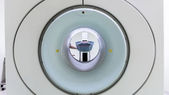
Advanced PET/MRI protocols boost lung cancer detection
PET/MRI achieves similar or superior results to PET/CT for the detection of pulmonary malignancies, according to new research [1].
Results of the meta-analysis were published on Jan. 24 in Radiology. Based on those findings, experts involved in the work indicate that MRI could have an increasingly prevalent role in lung cancer staging and restaging. Although previous research has resulted in findings somewhat conflicting on the modality’s efficacy for detecting small pulmonary lesions, this most recent study revealed that fluorine 18–labeled fluorodeoxyglucose (18F-FDG) PET/MRI performed as well as PET/CT in a cohort of nearly 1,300 individuals, even exceeding PET/CT detection results in some cases.
“Substituting the CT component with MRI has provided superior soft-tissue contrast and more functional information regarding lesion detection, resulting in a potentially higher diagnostic whole-body performance while simultaneously reducing patients’ radiation exposure,” study corresponding author Seyed Ali Mirshahvalad, from the Joint Department of Medical Imaging at Toronto General Hospital, and colleagues noted.
In total, the analysis included 43 studies involving 1,278 patients. The pooled sensitivity and specificity of 18F-FDG PET/MRI was comparable to 18F-FDG PET/CT, at 96% versus 99% and 100% versus 99%.
However, when advanced protocols involving contrast media and diffusion-weighted imaging were used, 18F-FDG PET/MRI outperformed CT in sensitivity.
The authors acknowledged that widespread access to MRI remains a limitation of the modality, as does cost-effectiveness. But, advances in MRI protocols appear to have overcome its previously documented limitations in detecting smaller neoplasms, the authors noted. They suggested that this could represent a growing role in lung cancer imaging for the modality.
“Even with the currently used PET/MRI protocols and their limitations, there may be a clinical role for 18F-FDG PET/MRI in lung cancer staging and restaging,” they noted, adding that current lung cancer imaging guidelines support a multimodality approach.
The study abstract is available here.

