Real-time MRI shows exact mechanisms underlying man's stutter
Real-time MR imaging could help experts fine-tune speech therapy for people who struggle with stuttering.
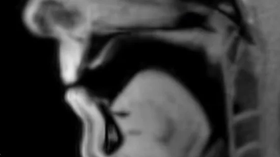
Researchers with the University Medical Center Göttingen (UMG) and the Max Planck Institute for Multidisciplinary Sciences (MPI-NAT) have recorded a live MRI scan of a man with a stutter reading a text message.
In the video, the man, who has had a stutter since childhood, can be heard struggling to get through certain parts of the text message. The muscle movements underlying his stutter can be seen at each point when he gets hung up on a word or sound, providing experts detailed new insight into the type of muscle malfunction that causes a person to stutter.
Prior to these developments in real-time imaging, identifying the mechanisms that cause altered speech patterns was difficult because the muscle involved in speech could not be fully visualized.
This work is the result of a collaboration between Professor Martin Sommer, leader of the Interdisciplinary Working Group for Fluency Disorders at the UMG, and the Biomedical NMR research group led by Professor Jens Frahm at the MPI-NAT.
Professor Sommer suggested that visualizing someone’s muscle movements in real-time as they stutter, more targeted therapies can be developed.
"By showing us the mechanical origin of the symptoms, real-time MRI improves our understanding of stuttering and will be an important tool in further research,” Sommer said in a release. “And by enabling us to directly see where the internal speech muscles and organs make ‘wrong’ movements, this method will also assist us greatly in the treatment of this multifaceted neuromuscular disorder."
The full results have been published in the Clinical Pictures section of the Lancet.
View the full video on YouTube.

