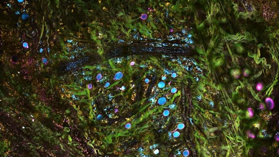
New microscope system may image cancer progression, tumorous cells in real time
A new molecular imaging system developed by researchers from the University of Illinois may allow researchers to monitor cancerous cells as they progress inside the body.
Simultaneous label-free auto fluorescence multi-harmonic (SLAM) microscopy utilizes tailored pulses of light to simultaneously image cancerous cells and tissue with multiple wavelengths, according to a University of Illinois news release published June 20.
“The way we have been removing, processing and staining tissue for diagnosing diseases has been practiced the same way for over a century,” said lead author Stephen Boppart, MD, PhD, a practicing physician and professor of bioengineering and electrical and computer engineering at the University of Illinois, in a prepared statement. “With advances in microscopy techniques such as ours, we hope to change the way we detect, visualize and monitor diseases that will lead to better diagnosis, treatments and outcomes.”
Using SLAM microscopy, Boppart and colleagues analyzed mammary tumors to see how they progressed.
"SLAM allows us to have a comprehensive picture of this ever-evolving tumor microenvironment at subcellular, molecular and metabolic levels in living animals and human tissue," Boppart said. "Monitoring that process can help us better understand cancer progression, and in the future could lead to better diagnosis of how advanced a tumor is, and better therapeutic approaches aimed at halting the progression.”
The researchers ultimately found healthy cells near the tumor had differences in metabolism and morphology—meaning the cells were “recruited” by the cancer. Additionally, SLAM microscopy allowed the team to see surrounding tissues creating infrastructure to support the tumor and observed communication between the tumor cells and surrounding cells in the form of vesicles.
Boppart's team is currently using SLAM microscopy to compare health tissue and cancer tissue in rats and humans, according to the news release, focusing on the relationship between vesicle activity and cancer aggressiveness.

