Novel PET method improves detection of prostate cancer recurrence
Prostate-specific membrane antigen (PSMA) PET/CT can better detect recurrent prostate cancer post-surgery compared to fluciclovine PET/CT, according to results of a July 30 study published in The Lancet Oncology.
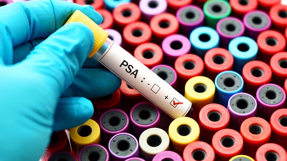
Following prostate cancer surgery, patients receive a blood test to check for prostate-specific antigen. A positive test indicates a recurrence of cancer and the patient typically undergoes fluciclovine PET/CT imaging—the current standard of care—to determine the exact location of the disease.
“However, another new imaging method exists that is called prostate-specific membrane antigen imaging, or PSMA PET/CT, that can also be used for the same purposes,” wrote Duane Bates, of UCLA Jonsson Comprehensive Cancer Center, in a UCLA news brief. “This scan is currently investigational and has not yet been approved by the FDA.”
In the prospective study, Jeremie Calais, MD, with UCLA’s Department of Molecular and Medical Pharmacology, and colleagues investigated whether 18F-fluciclovine or PSMA PET-CT scans could improve the localization of biochemical recurrence of prostate cancer in 50 patients between Feb. 26, 2018 and Sept. 20, 2018.
All men had prior radical prostatectomy, low PSA levels and received a second PSMA imaging scan within 15 days before or after their fluciclovine exam. All the images were read three times by three independent physicians.
Detection rates were much lower on 18F-fluciclovine scans compared to PSMA PET/CT, the authors noted. Using the latter molecular imaging technique, prostate cancer recurrence was found in 56% of scans compared to 26% of fluciclovine scans
“Prostate-specific membrane antigen imaging should be the positron emission tomography imaging agent of choice for men who have a prostate cancer recurrence after radical prostatectomy, which needs to be detected and located,” Bates wrote in the same brief. “For these men, it should become the standard of care.”

