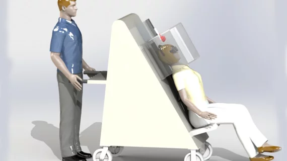
Researchers receive grant worth millions to develop portable PET scanner
Researchers at Weill Cornell Medicine just received a grant worth more than $6 million to develop neuroimaging technology that could expand access to Alzheimer's diagnostics.
The grant was gifted by the National Institute on Aging—part of the National Institutes of Health—to fund the building of a portable, upright PET scanner that will enable the exams to be done while patients sit down. This could increase access in areas where PET scans are not routinely available.
“We can move it to medical centers that might not have advanced brain imaging, enabling us to provide the highest-level care to more diverse populations,” Amir H. Goldan, PhD, an associate professor of electrical engineering in radiology at Weill Cornell Medicine, said in a release.
Goldan and colleagues previously developed a PET scanner said to have the world’s highest resolution. That scanner—dubbed Prism-PET—is capable of detecting some of the smallest areas of radiotracer concentrations on imaging, and it does so in great detail.
The director of the Brain Health Imaging Institute at Weill Cornell Medicine, Gloria Chiang, will also be involved in the project. Chiang and Goldan are going to focus their efforts on homing in Prism-PET's ability to detect tau tangles in the transentorhinal cortex.
This area of the brain is known to be associated with memory and perception of time. It also happens to be a location where some of the earliest signs of tau tangles develop, making it an area of great interest in research on the progression of Alzheimer’s disease. Its experts are able to identify these areas on imaging earlier—even before patients start to show signs of cognitive decline—providers could potentially initiate therapeutic interventions to slow the progression of symptoms.
The transentorhinal cortex can be difficult to see in detail on imaging, Chiang explained. The hope is that the new scanner could improve the visualization of this area.
The grant also follows the recent approval of lecanemab—a monoclonal antibody that targets amyloid plaques and has been shown to slow the progression of symptoms related to Alzheimer's. Patients who are given the drug require routine imaging to track their progress on the medication, and to monitor for signs of adverse events. But access to these scans can be hard to come by for many. A portable scanner could help address these barriers.
“Our overall goal is two-fold—having the highest performance brain PET scanner available, while also addressing its accessibility and portability to serve the community,” Goldan said. “The earlier you can diagnose Alzheimer's disease, the better the chances of therapy being effective.”
It is estimated that up to 6.7 million people 65 and older in the United States are living with Alzheimer’s today. In 2023, the Alzheimer’s Association cautioned that, without effective treatments, this figure has the potential to double in the coming decades.
Learn more about the project here.

