MRI affirms early HIV treatment key to prevent neurological damage
Although scientists have known for some time about how the human immunodeficiency virus (HIV) negatively effects the brain, it remains uncertain when these changes start and how combination antiretroviral therapy (cART) stops or slows its progression.
However, in a collaboration between researchers from McGill University in Canada, the Washington University in St. Louis and Yale University, MRI brain data have revealed that early HIV treatment is essential for patients to avoid neurological damage caused by the virus, according to a May 3 press release from McGill University.
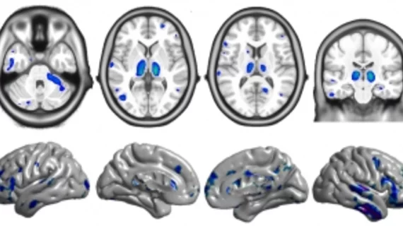
Specially, researchers found that the longer a patient with HIV goes untreated, the greater the volume loss and cortical thinning in several regions of the brain.
The research was published online in the journal Clinical Infectious Diseases on April 24.
“There have been few longitudinal structural neuroimaging studies in early HIV infection, and none that have used such sensitive analysis methods in a relatively large sample,” said lead author Ryan Sanford, a PhD candidate at McGill, in a prepared statement. “The findings make the neurological case for early treatment initiation and sends a hopeful message to people living with HIV that starting and adhering to cART may protect the brain from further injury.”
The researchers analyzed brain MRI data from 65 patients at the University of California San Francisco who had been infected with HIV less than a year prior. The data was then compared to the MRI data of 19 HIV-negative participants and 16 HIV-positive patients who had been infected with HIV for a minimum of three years.
HIV-positive participants treated with combination antiretroviral therapy (cART) were found to have maintained volume in the brain and increased cortical thickness in the frontal and temporal lobes, according to the press release.
“It [study findings] also helps focus the search for possible mechanisms of brain injury in this condition, pointing towards injury that occurs principally during untreated infection," Sanford added. "This opens the door to novel treatments focused on potentially reversing these observed structural changes.”

