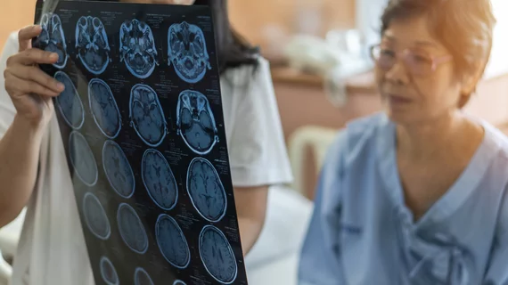
Expansion of open-source neuroimaging dataset aims to boost stroke research
Researchers recently revamped an open-source neuroimaging database in the hopes of expanding algorithm development in the field of stroke care.
With accurate lesion segmentation in mind, the USC-led expansion built upon the Anatomical Tracings of Lesion After Stroke (ATLAS), version 1.2. ATLAS 1.2 contained hundreds of stroke T1w MRIs and manually segmented lesion masks, and though the intentions were good the results were less so. Many algorithms developed using that dataset did not yield acceptable accuracy or were improperly invalidated, limiting the usefulness of the data in stroke research.
To compensate for this, researchers expanded upon the original data, curating a larger dataset of T1w MRIs and manually segmented lesion masks—ATLAS 2.0.
Citing the time-consuming nature of manual lesion segmentation, lead author of the study Sook-Lei Liew, PhD, of the Mark and Mary Stevens Neuroimaging and Informatics Institute, Keck School of Medicine at the University of Southern California, Los Angeles, and colleagues elaborated on how the data expansion can improve stroke research:
“Algorithm development using this larger sample should lead to more robust solutions; the hidden datasets allow for unbiased performance evaluation via segmentation challenges. We anticipate that ATLAS v2.0 will lead to improved algorithms, facilitating large-scale stroke research.”
Developing ATLAS 2.0 was a collaborative effort shared by 16 co-authors across the university. It contains 1,271 manually segmented lesions and has already been downloaded by thousands of researchers across the world, according to the announcement shared by Keck School of Medicine of USC.
“With the progression of this work, we hope to see a future where clinicians can analyze an individual’s data to discover their likelihood of responding to different treatments. Using a precision medicine approach, their stroke rehabilitation therapy could be personalized to maximize their recovery,” Liew said.
To learn how to access the ATLAS data, refer to USC Stevens Neuroimaging and Informatics Institute.
More details about the data can be viewed here.
Related neuroimaging news:
We can now diagnose Alzheimer's using only an MRI and an algorithm
NIH grants $3.5 million toward researching COVID's neurological impact
New 7T MRI scanners could increase radiologists' role in treating neurological conditions
Stroke care still lags among certain Medicare populations

