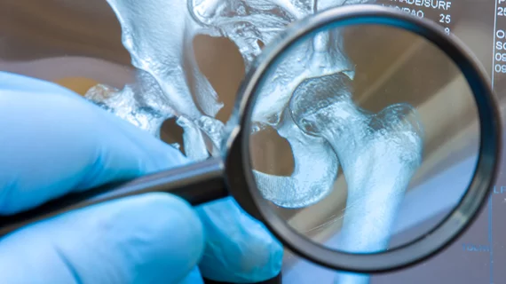
Diagnosing osteoporosis using a deep-radiomics-based approach
Researchers have developed deep-radiomics-based models capable of predicting osteoporosis with high accuracy and without need for DXA imaging.
Their work was published recently in Radiology: Artificial Intelligence. In the paper, experts detail how they extracted radiomics features from nearly 5,000 hip radiographs, which were used to train multiple models to detect minute signs of osteoporosis.
Although bone mineral density scans are the reference standard for diagnosing osteoporosis, they continue to be underutilized and may not be available at all in lesser developed countries, authors of the study pointed out.
“Its use for screening and posttreatment follow-up in regions with poor economies is limited due to the low availability of scanners and relatively high cost. Even in developed countries, many patients are left at risk for fracture without undergoing osteoporosis screening due to lack of understanding among health care providers,” corresponding author Hee Dong Chae, from the Department of Radiology at Seoul National University Hospital, and co-authors explained.
For these reasons, the utility of deep learning radiomics-based approaches to assist in diagnosis using routine radiographs has been explored. Though promising, studies pertaining to this approach have been limited in their feature extractions and comparisons.
For this study, researchers used a multitude of feature extractions to train the deep-radiomics-based models—10 deep features, 16 texture features and three clinical features. In total, seven models were developed, each of which used different combinations of features to diagnose osteoporosis. Area under the receiver operating characteristic curve (AUC) was used grade diagnostic performance and 6 radiologist completed observer agreement tests.
Each model performed well, but the model that included deep, texture and clinical features yielded the best results, with an AUC of .95 and an observer performance of .87, respectively.
“Deep-radiomics model using hip radiographs can diagnose osteoporosis with high performance. Our model can serve as an opportunistic diagnostic test to triage patients at risk for osteoporosis for further confirmatory tests, including DXA.” the authors concluded.
More on AI and radiomics:
Deep learning algorithm predicts emphysema mortality
DL networks augment radiologist performance for thyroid cancer detection
AI-based mammo screening protocol reduces radiologist workload by 62%
MRI-based radiomics boosts triple-negative breast cancer detection
References:
Deep-Radiomics-Based Approach to the Diagnosis of Osteoporosis Using Hip Radiographs, Sangwook Kim, Bo Ram Kim, Hee-Dong Chae, Jimin Lee, Sung-Joon Ye, Dong Hyun Kim, Sung Hwan Hong, Ja-Young Choi, and Hye Jin Yoo. Radiology: Artificial Intelligence

