
PHOTO GALLERY: Abdominal, pelvic and rectal foreign bodies
This is a clinical photo gallery showcasing the uncommon findings of foreign bodies in pelvic and abdominal imaging, including rectal foreign bodies (RFB). The collection includes images from computed tomography and X-ray. Most of these images include items patients inserted into their own bodies.
RFBs are more common than many people may think. Some of the studies where these images came from said the incidents are on the rise in emergency rooms. A children's hospitals saw enough of these cases that it posted clinical images on social media to warn patients and parents. A hospital in Germany said in a 10-year study of its ER records, they had 22 RFB presentations in 20 patients.
Hover over the images to view captions, and click on them to enlarge.
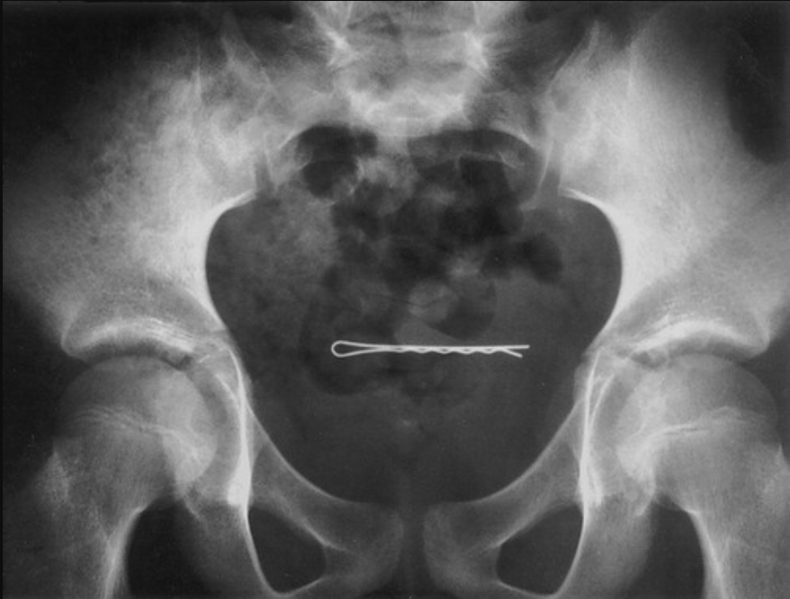
A 19-year-old woman with a bobby pin lodged in her uterus from an attempted abortion. Image courtesy of RSNA
A 51-year-old man with a bottle of gargling liquid in a Hartman pouch from earlier colon surgery to remove an impacted shampoo bottle. Image courtesy of RSNA
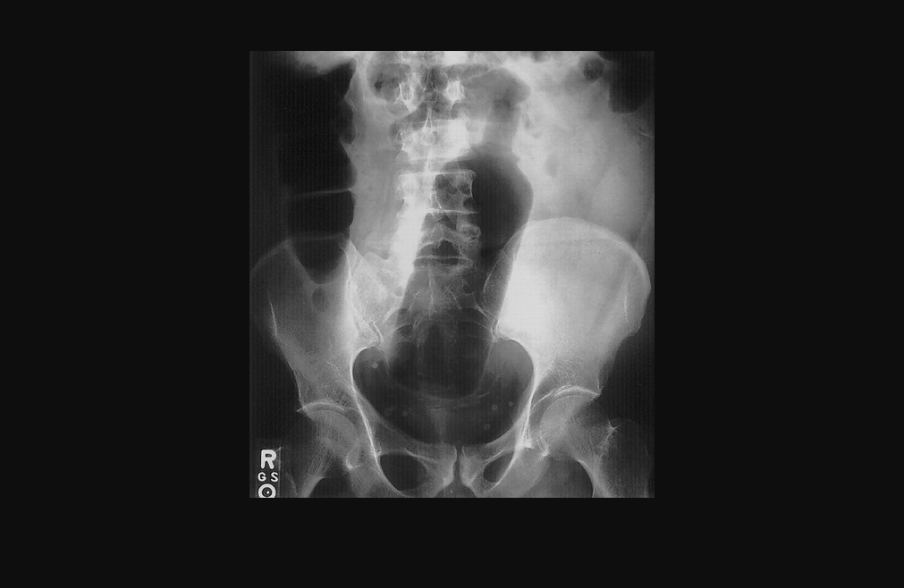
Radiograph of a 60-year-old man with a condensed milk can that he inserted into his rectum. Image courtesy of RSNA
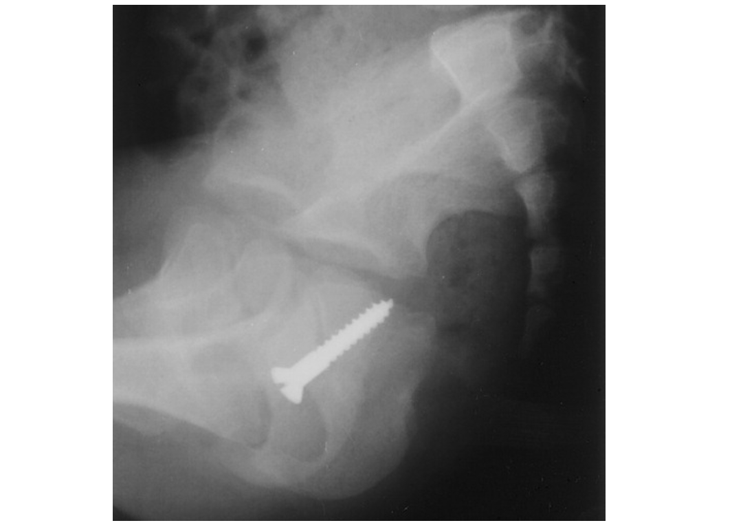
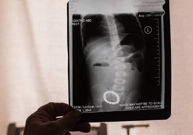
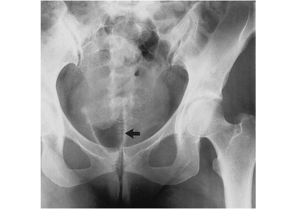
Radiograph of a young child shows a screw that she had inserted into her vagina. Image courtesy of RSNA
Smuggled narcotics packet on contrast-enhanced CT scan. Image courtesy of RSNA
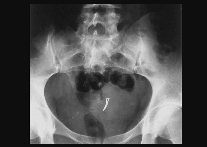
X-ray of a 2-year-old girl shows a bobby pin in her bladder. Image courtesy of RSNA
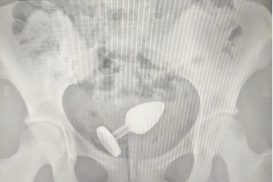
An anal plug sex toy retained in the colon. The children's hospital that captured this image said it is not uncommon for children, especially teenagers, to come into the emergency department with items inserted into their rectums. Photo courtesy of Okhmatdyt Children's Hospital, Kyiv, Ukraine.
A perfume bottle retained in colon after a teenager inserted it into their rectum. Okhmatdyt Children's Hospital in Kyiv, Ukraine, posted this with other images as a public education social media post because the hospital removes these types of items on a regular basis. They said it is not uncommon for children, especially teenagers, to come into the emergency department with items inserted into their rectums. Photo courtesy of Okhmatdyt Children's Hospital
A perfume bottle retained in colon after a teenager inserted it into their rectum. Okhmatdyt Children's Hospital in Kyiv, Ukraine, posted this with other images as a public education social media post because the hospital removes these types of items on a regular basis. They said it is not uncommon for children, especially teenagers, to come into the emergency department with items inserted into their rectums. Photo courtesy of Okhmatdyt Children's Hospital
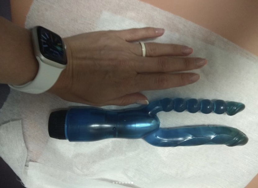
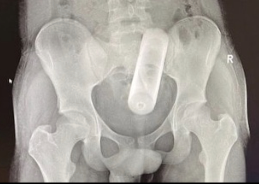
A surgically removed vibrator that was retained in the colon after a teenager inserted it into their rectum. Courtesy of Okhmatdyt Children's Hospital Kyiv, Ukraine.
The same vibrator retained in the colon on X-ray. Okhmatdyt Children's Hospital in Kyiv, Ukraine, posted this with other images as a public service message on social media post because the hospital removes these types of items on a regular basis. Courtesy of Okhmatdyt Children's Hospital.
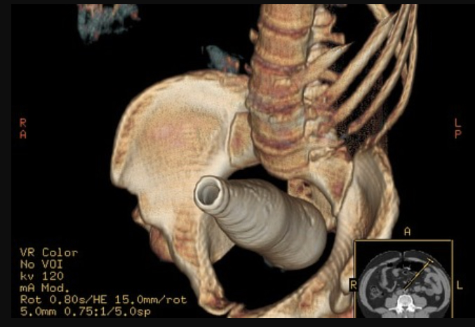
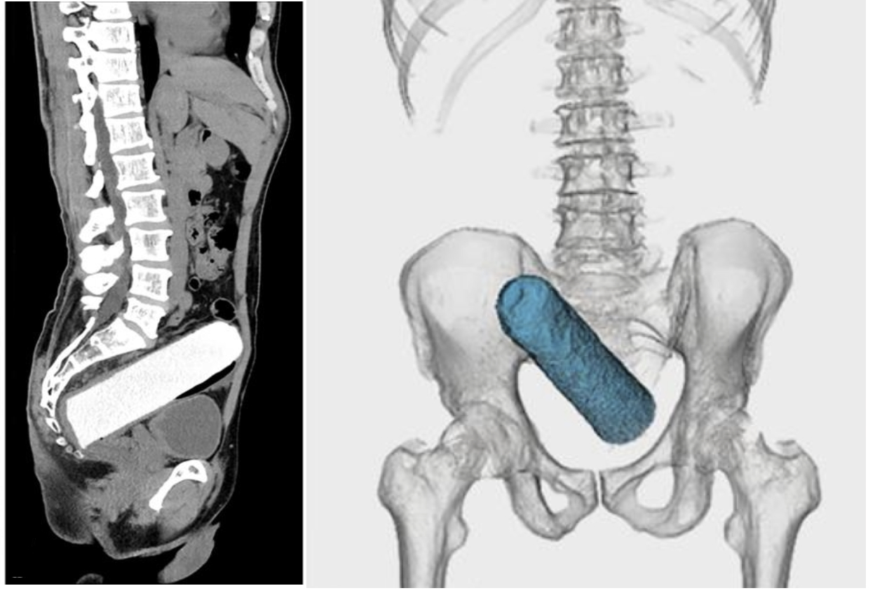
A 3D reconstructed CT scan image of a Cola-Cola bottle projecting over the pelvis within the rectum and distal sigmoid colon. Ghonaim E, Rectal foreign body. Case study, Radiopaedia.org
Axial CT scan of a radiodense foreign body (coca cola bottle) within the rectum and distal sigmoid colon. Ghonaim E, Rectal foreign body. Case study, Radiopaedia.org
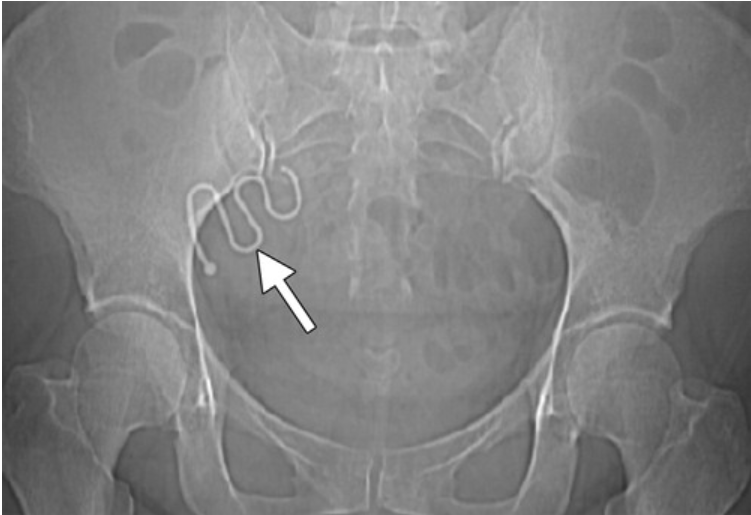
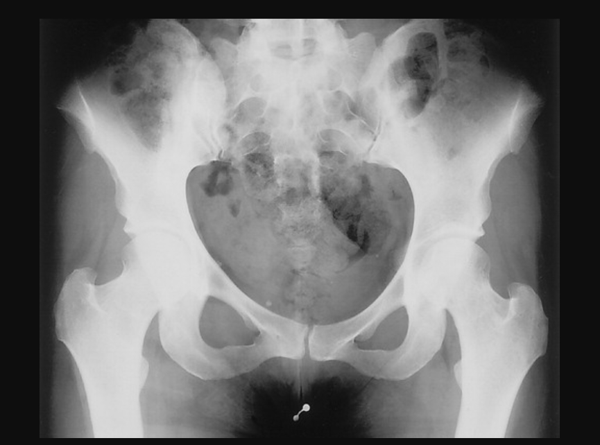
Radiograph of an 18-year-old man demonstrates bladder calculi that had formed on fine telephone wire. Six months before, he lost the wire in his bladder when he achieved an erection while inserting the wire into his urethra during masturbation. Image courtesy of RSNA
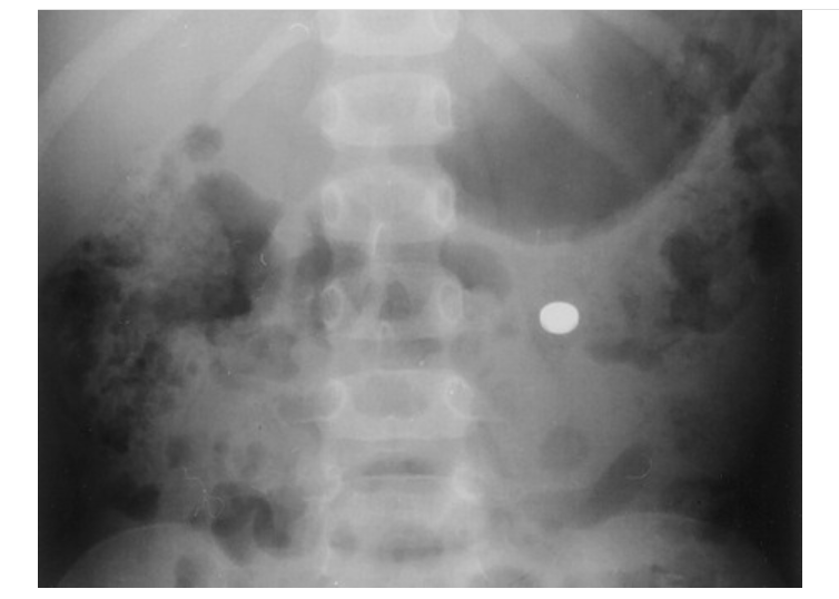
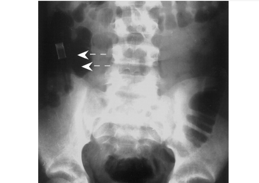
A watch battery in the transverse colon. It was ingested by a 4-year-old girl and passed without complications. Image courtesy of RSNA
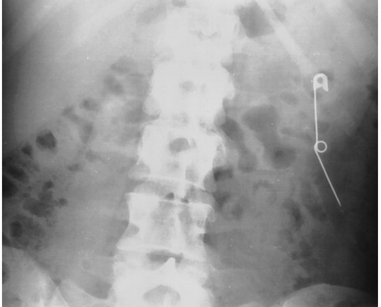
X-ray of a 28-year-old woman who periodically swallowed pins and razor blades shows an open safety pin in her descending colon. The pin passed without difficulty. Courtesy of RSNA.
An intrauterine device (IUD) migration on a CT topogram helps identify the object (arrow) as a migrated IUD. Courtesy of RSNA https://pubs.rsna.org/doi/full/10.1148/rg.312105123
Radiograph shows a pencil (arrows) lodged in the ascending colon. It passed without difficulty, which is unusual for such an elongated object to pass through the intestinal tract without problems. Courtesy of RSNA https://pubs.rsna.org/doi/10.1148/rg.233025137
An intrauterine device (IUD) migration seen on an axial CT scan (arrow). The device is more apparent and easier to identify using the CT scout scan. Courtesy of RSNA https://pubs.rsna.org/doi/full/10.1148/rg.312105123
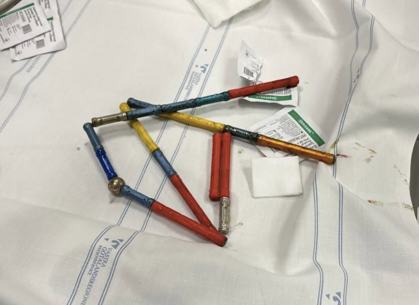
A child swallowed 20 magnets that remained connected through different parts of the stomach and intestinal tract. Doctors managed to grab the first magnet and pull it out along with a chain of 18 elements. Surgeons had to remove last one. Photos courtesy of Okhmatdyt Children's Hospital, Kyiv, Ukraine.
A child swallowed 20 magnets that remained connected through different parts of the stomach and intestinal tract. Doctors managed to grab the first magnet and pull it out along with a chain of 18 elements. Surgeons had to remove last one. Photos courtesy of Okhmatdyt Children's Hospital, Kyiv, Ukraine.
X-ray of neodymium magnets swallowed and in the stomach of a baby. These powerful magnets can pull through the walls and cause holes in all organs. In most cases, such as this one, the child needs immediate surgery. Photos courtesy of Okhmatdyt Children's Hospital, Kyiv, Ukraine.
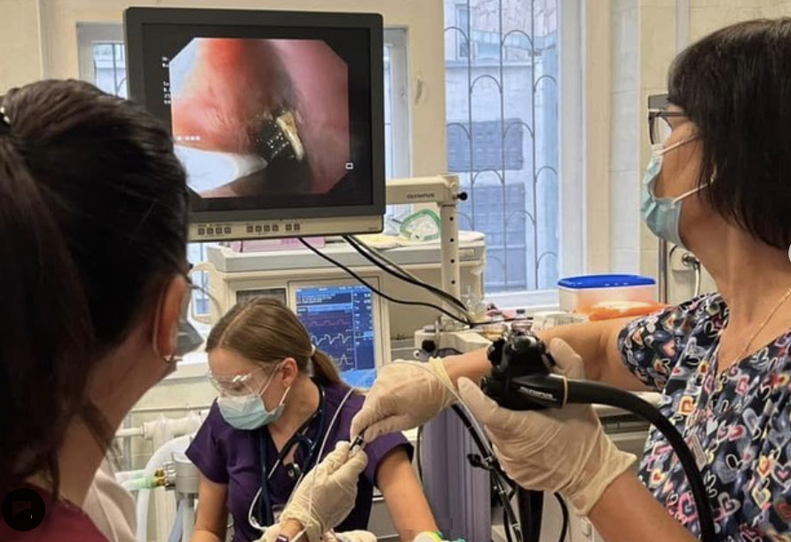
Physicians extracting a 19 cm toothbrush from the esophagus of a teenager who swallowed it by accident when trying to cause vomiting after eating in an attempt to lose weight. The hospital said they remove a few tooth brushes each year for this reason. Photos courtesy of Okhmatdyt Children's Hospital, Kyiv, Ukraine.
A 33-year-old man from long-term psychiatric facility who ingested multiple foreign bodies preoperative coronal CT multiplanar reconstruction identified numerous gastric foreign bodies. Note that plastic ballpoint pen (black arrow) is radiolucent, whereas USB cable is radiopaque (white arrow). Multiple disposable plastic spoons (arranged to right of ballpoint pen) are radiolucent and not visualized on CT. Image courtesy of AJR
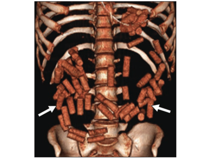
A 27-year-old man from a Central American country presented with vomiting. More than 40 drug containers were surgically removed. 3D volume-rendered image showing multiple drug-containing radiopaque packets. Image courtesy of AJR
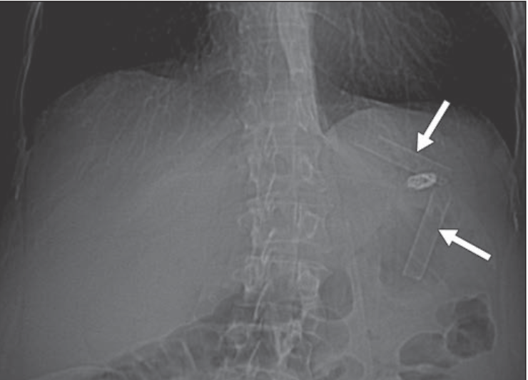
A 47-year-old woman with history of drug abuse who swallowed a crack cocaine glass pipe in two pieces to avoid police arrest.
CT scout image shows two glass tubular fragments (arrows) in stomach. Only one piece of glass was retrieved from the gastric body during endoscopy. The other piece was followed with radiographs until elimination. Image courtesy of AJR
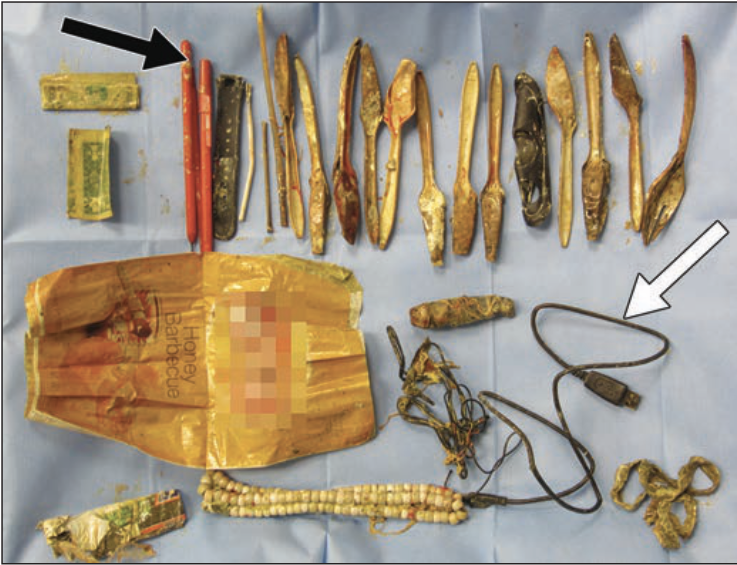
From a 33-year-old man from a long-term psychiatric facility who ingested multiple foreign bodies. Preoperative coronal CT multiplanar reconstruction identified numerous gastric foreign bodies. Note that plastic ballpoint pen (black arrow) is radiolucent, whereas USB cable is radiopaque (white arrow). Multiple disposable plastic spoons (arranged to right of ballpoint pen) are radiolucent and not visualized on CT. Image courtesy of AJR
Scout radiograph readily identifies piece of glass (arrow) in pelvis. Object is obscured by oral contrast agent when viewed in soft-tissue windows. Image courtesy of AJR
Scout radiograph readily identifies piece of glass (arrow) in pelvis. Object is obscured by oral contrast agent when viewed in soft-tissue windows. Image courtesy of AJR
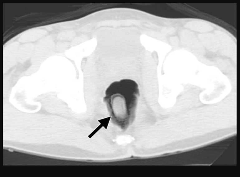
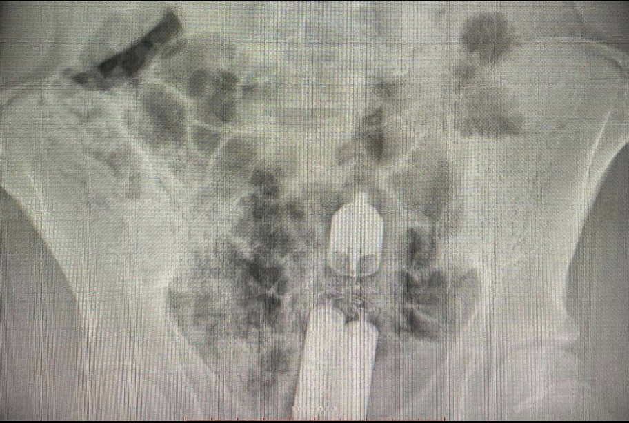
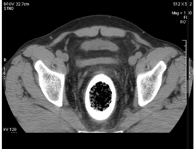
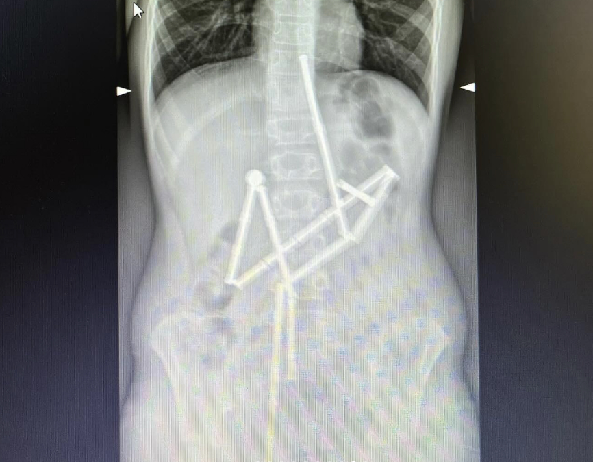
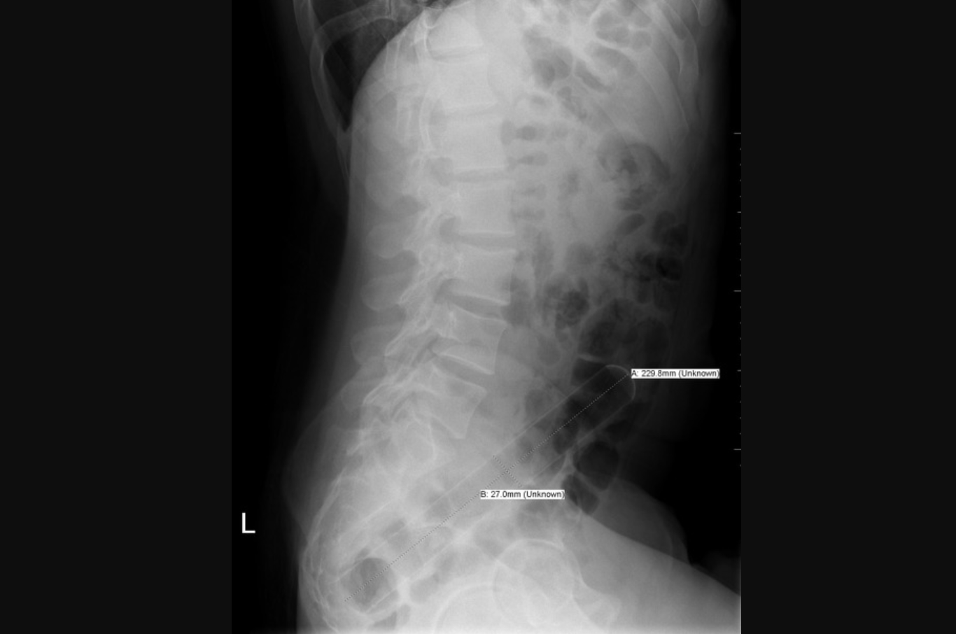
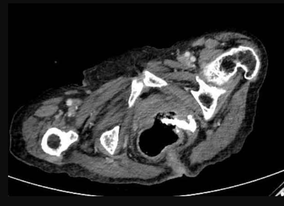
Case from Japan. Abdominal CT imaging views of a silicone foreign body inserted it into the rectum (patient was unable to retrieve it). X-ray fluoroscopy showing the lateral pelvic view of the foreign body in a rectosigmoid position. They changed the axis of the foreign body by manual abdominal compression and moved it to the lower rectum where it was removed by manual transanal extraction. Sigmoidoscopy following extraction showed mild erosion in the rectal mucosa, but no findings of bleeding or perforation. Images courtesy of Case Reports in Gastroenterology
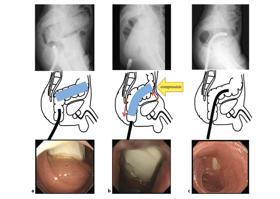
Case from Japan. Abdominal CT imaging shows a silicone foreign body inserted it into the rectum. X-ray fluoroscopy showing the lateral pelvic view of the foreign body in a rectosigmoid position. They changed the axis of the foreign body by manual abdominal compression and moved it to the lower rectum where it was removed by manual transanal extraction. Sigmoidoscopy following extraction showed mild erosion in the rectal mucosa, but no findings of bleeding or perforation. Images courtesy of Case Reports in Gastroenterology
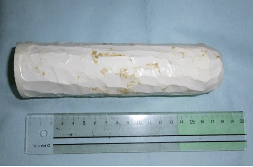
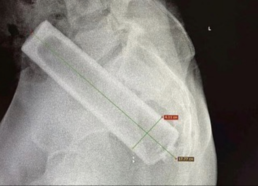
Case from Japan. The silicone foreign body, made by the patient himself, that was inserted it into the rectum and unable to be retrieved. After it was removed by manual extraction, the item measured 18 x 4 cm. Images courtesy of Case Reports in Gastroenterology
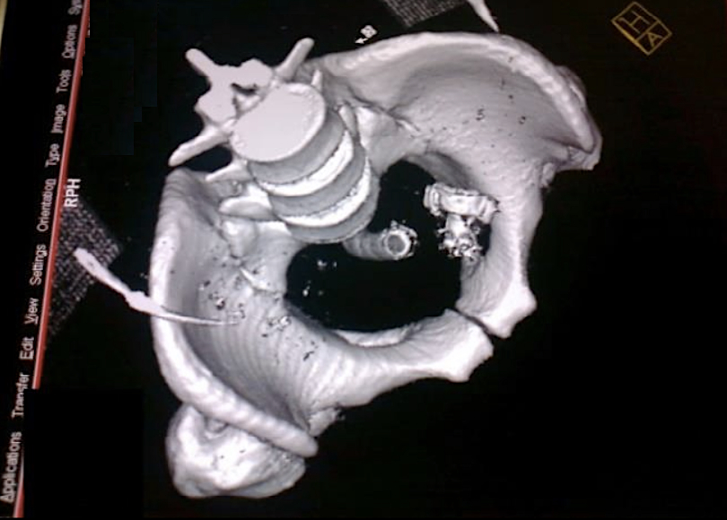
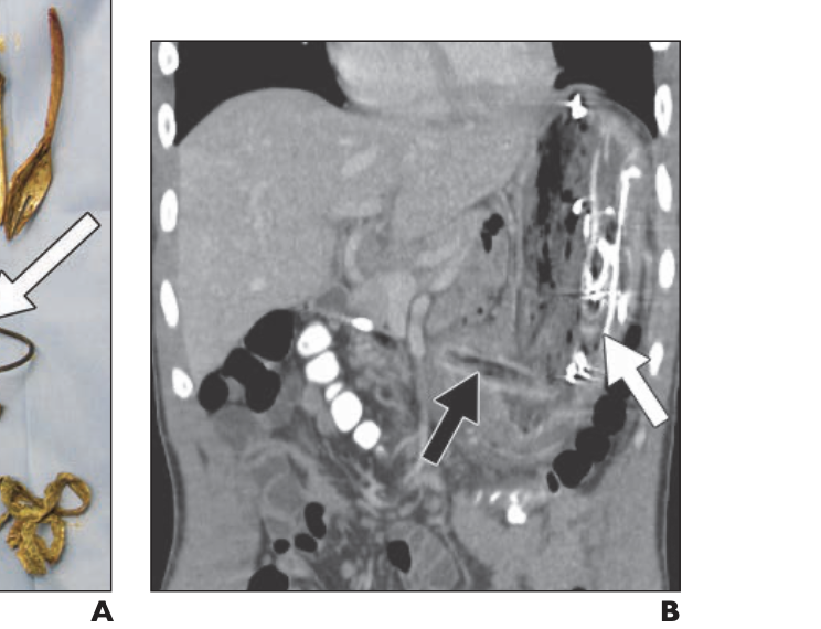
Case from India. A 22-year-old male inserted a hand-held bidet shower head with 8 cm of hose attached into his rectum. Physicians were unable to extract the object transanally under anesthesia due to the handle of the object being entangled in the bowel mucosa, so a laparotomy was performed. Courtesy of International Surgery Journal.
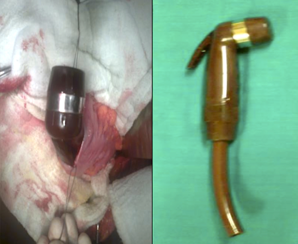
Case from India. A 22-year-old male inserted a hand-held bidet shower head with 8 cm of hose attached into his rectum. Physicians were unable to extract the rectal foreign object transanally under anesthesia due to the handle of the object being entangled in the bowel mucosa, so a laparotomy was performed. Courtesy of International Surgery Journal.
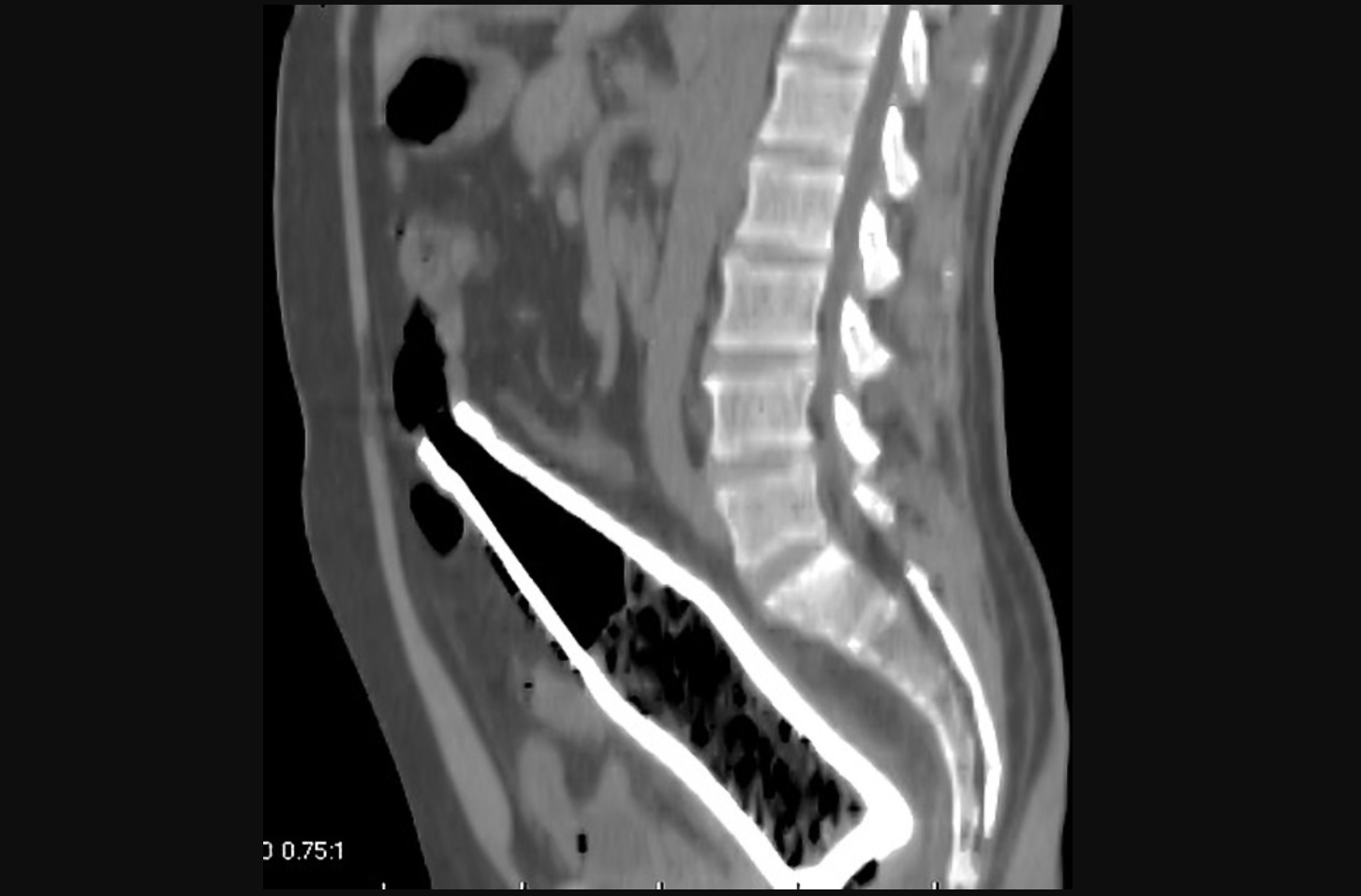
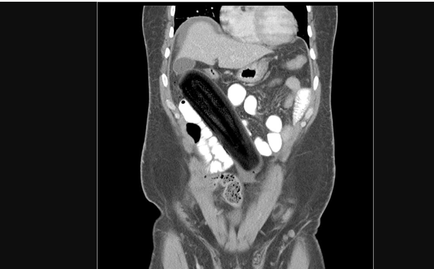
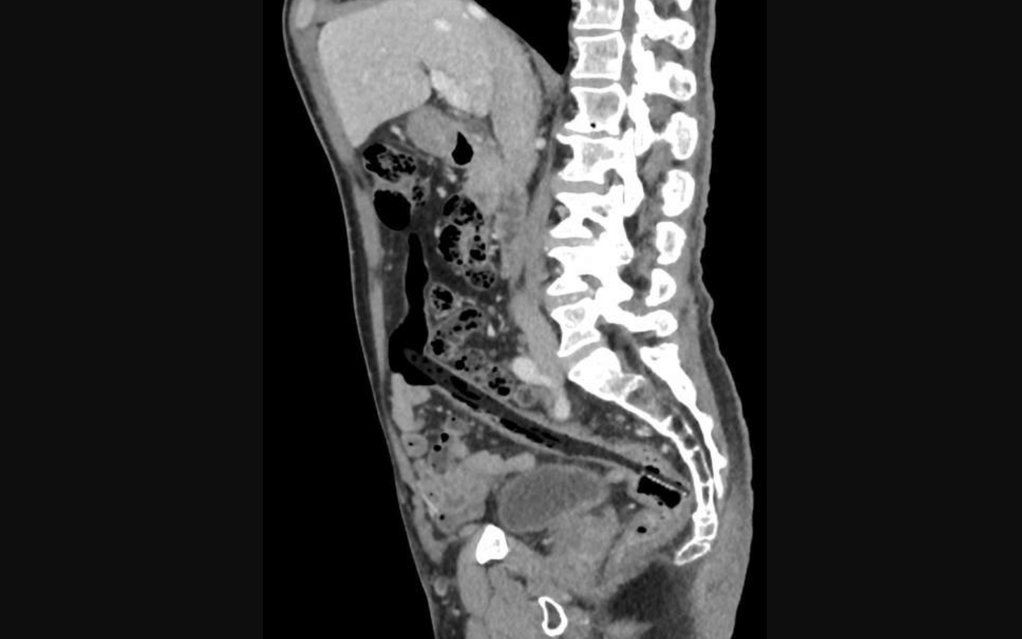
Sagittal CT image of a Coca-Cola bottle within the rectum and distal sigmoid colon. Ghonaim E, Rectal foreign body. Case study, Radiopaedia.org
Radiograph of an adult smuggling drugs shows relatively opaque packets in the transverse and descending colon. Image courtesy of RSNA
Radiograph of an elderly woman shows a menstrual cup (arrow) that she had inserted into her vagina. Image courtesy of RSNA
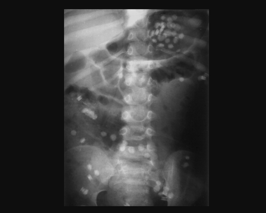
Radiograph of a 3-year-old boy showing multiple iron tablets, probably containing ferrous gluconate and ferrous sulphate salts. He recovered without sequelae. Image courtesy of RSNA
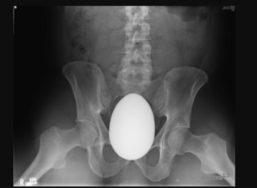
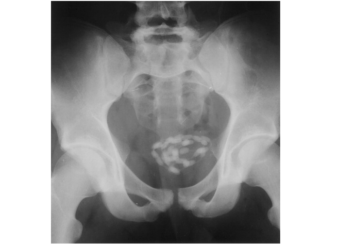
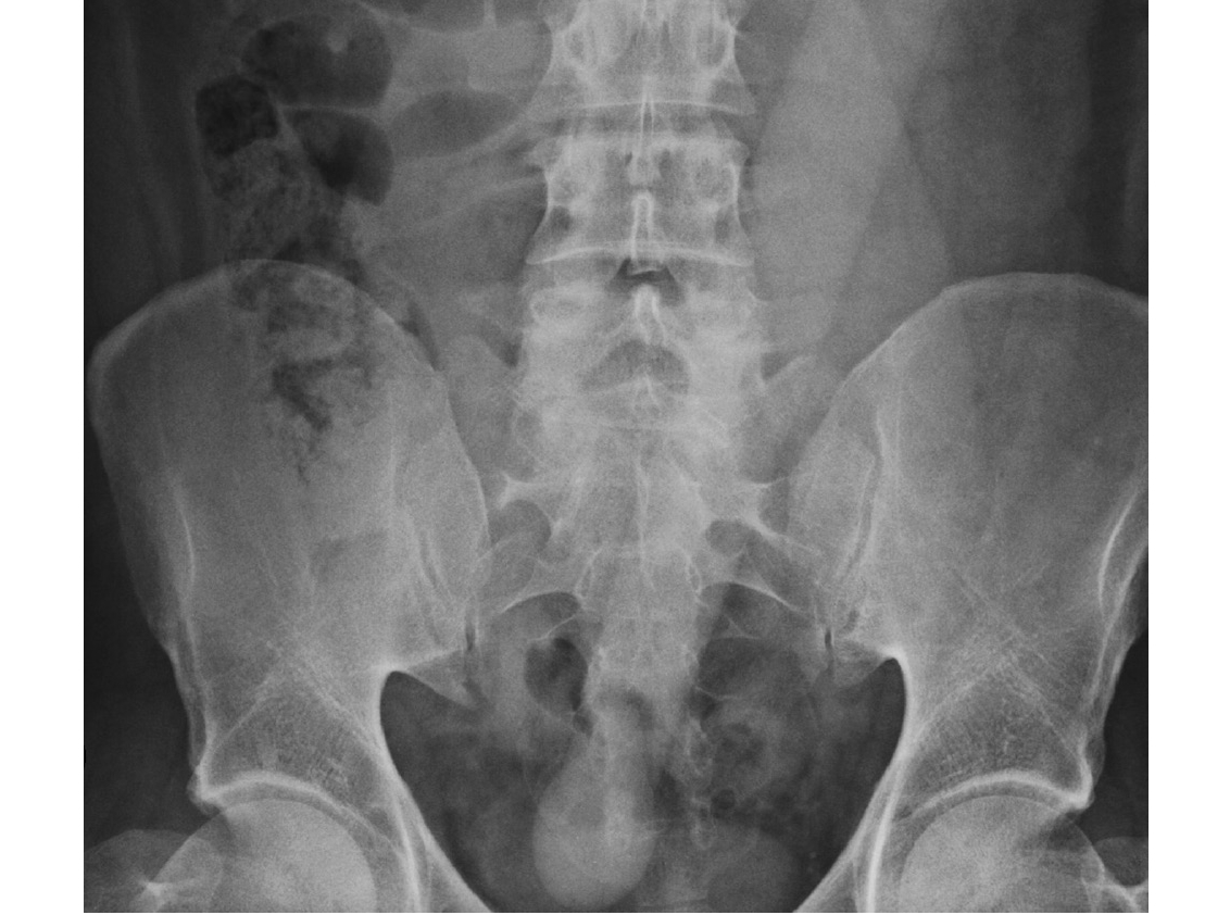
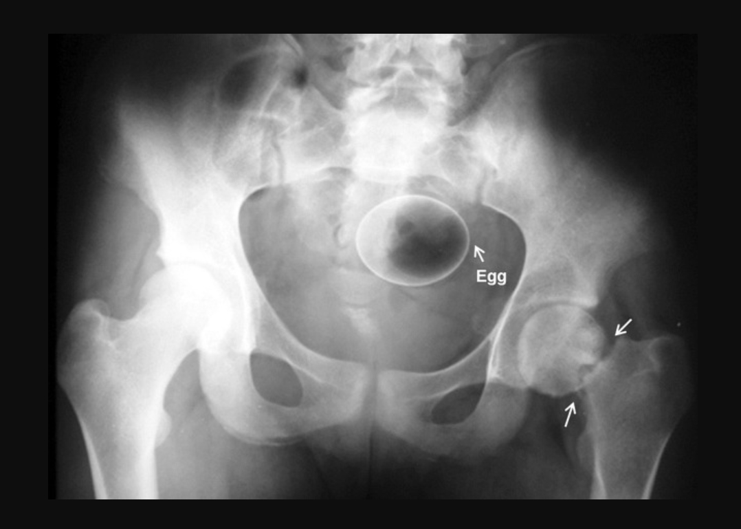
A 41-year-old male presented to the emergency department with a marble egg inserted and tightly wedged in the pelvic anatomy of the the distal sigmoid and rectum. It required surgery to remove the egg-shaped stone rectal foreign body. Image courtesy of the International Journal of Surgery Case Reports.
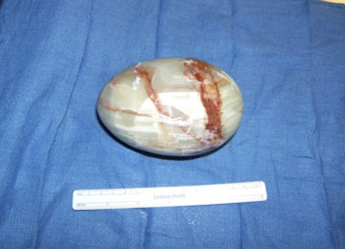
A 41-year-old male presented to the emergency department an marble egg inserted and tightly wedged in the pelvic anatomy of the the distal sigmoid and rectum. It required surgery to remove the egg-shaped stone rectal foreign body. Image courtesy of the International Journal of Surgery Case Reports.
After rectal perforation, free-lying dildo in the abdomen. The case is from a center in Germany that reported its experience with 22 emergency room visits from 20 patients with RFB between 2006 and 2016. Courtesy of the journal Innovative Surgical Sciences.
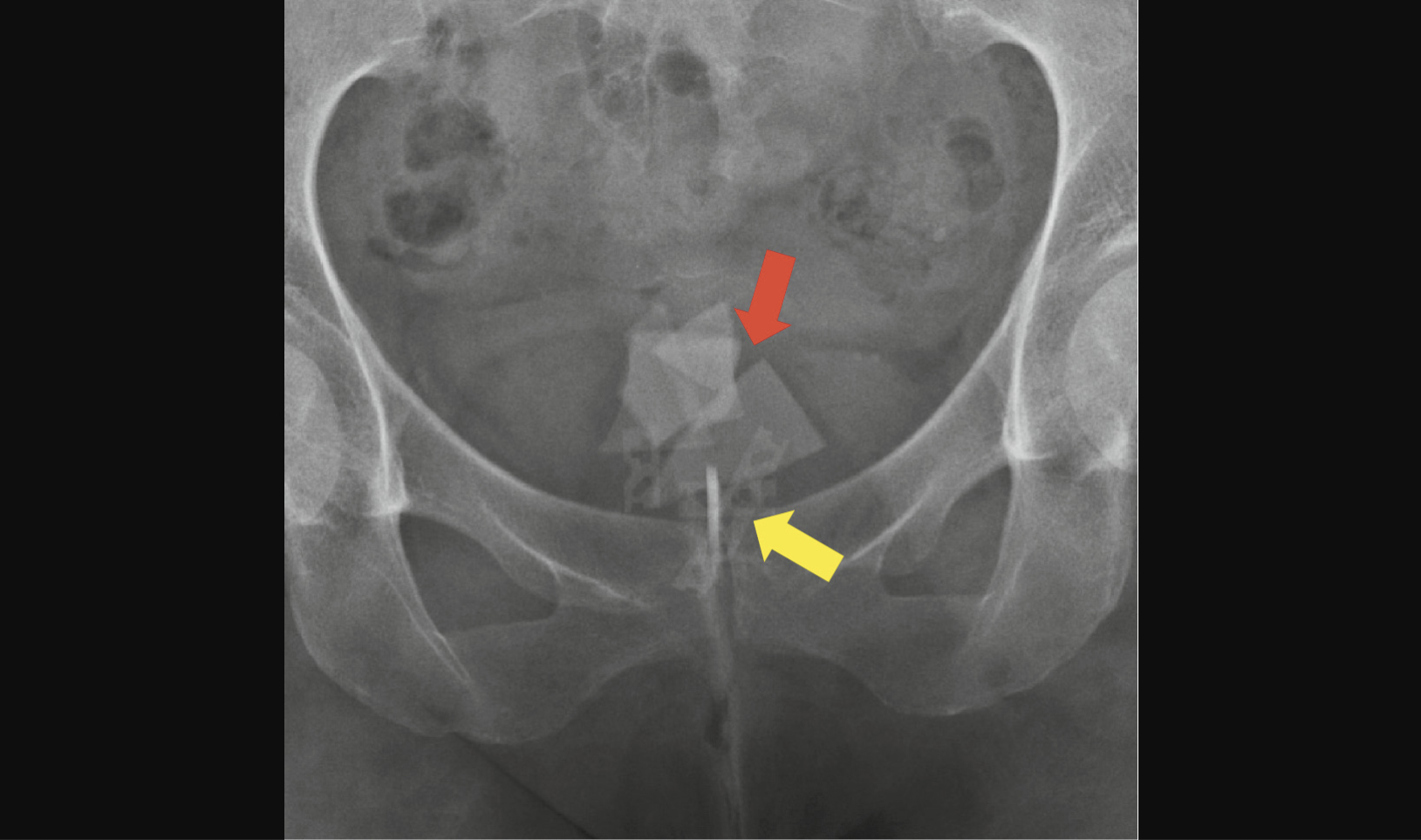
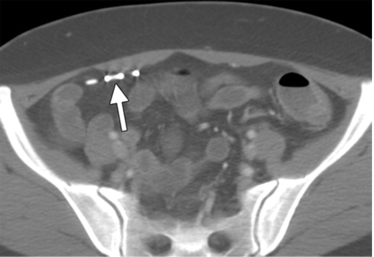
Peranally and transvaginally inserted glass fragments (red arrow) and razor blades (yellow arrow). The case is from a center in Germany that reported its experience with 22 emergency room visits from 20 patients with RFB between 2006 and 2016. Courtesy of the journal Innovative Surgical Sciences.
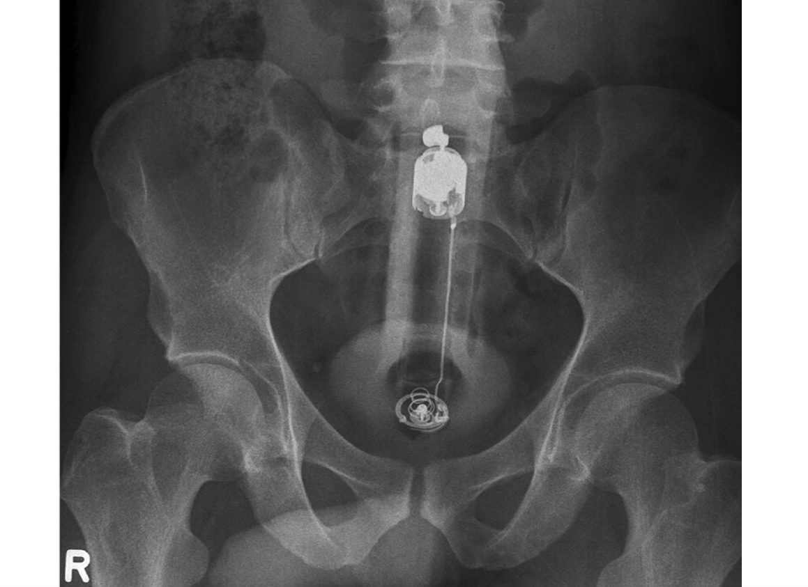
Complete peranally inserted vibrator. The case is from a center in Germany that reported its experience with 22 emergency room visits from 20 patients with RFB between 2006 and 2016. Courtesy of the journal Innovative Surgical Sciences.
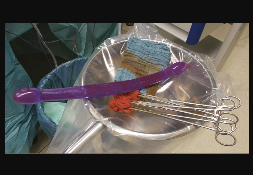
This 40-cm-long dildo that perforated a patient's sigmoid and had to be removed surgically from the abdomen. The case is from a center in Germany that reported its experience with 22 emergency room visits from 20 patients with RFB between 2006 and 2016. Courtesy of the journal Innovative Surgical Sciences.
Radiograph of a young woman showing a labial ring. Image courtesy of RSNA
Aluminum cigar cover tube foreign body in the rectum of a patient. Aluminum is a low attenuation metal compared to other metals in X-ray, so it is barely visible. Image courtesy of Radiopaedia
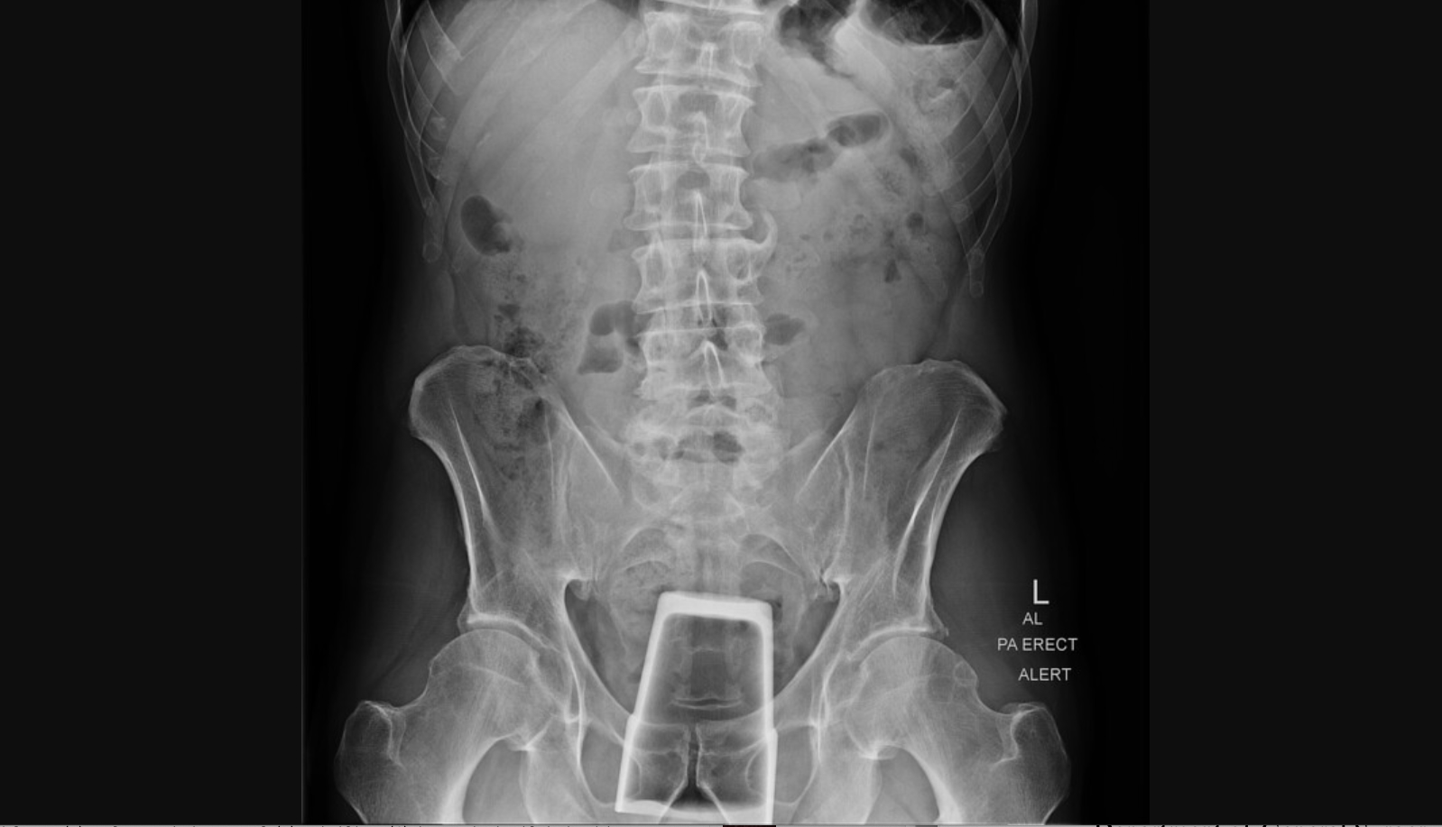
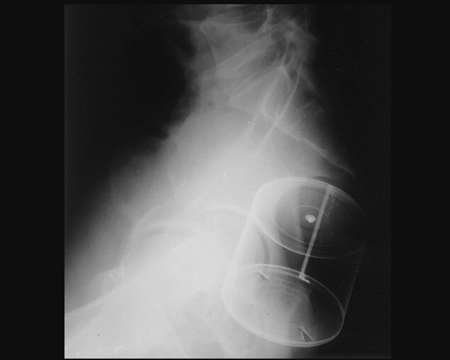
Drinking glass tumbler inserted into rectum. Image courtesy of Radiopaedia
An egg inserted into the rectum. Image courtesy of Radiopaedia
Eggplant inserted into rectum. Image courtesy of Radiopaedia
Rectovagino-vesical fistula caused by dentures. Dentures are sometimes swallowed by the elderly and can move through the gastrointestinal tract. Image courtesy of Radiopaedia
Zucchini inserted into the rectum. Image courtesy of Radiopaedia
Tooth brush inserted into the rectum. Image courtesy of Radiopaedia
Related Content:

Dave Fornell has covered healthcare for more than 17 years, with a focus in cardiology and radiology. Fornell is a 5-time winner of a Jesse H. Neal Award, the most prestigious editorial honors in the field of specialized journalism. The wins included best technical content, best use of social media and best COVID-19 coverage. Fornell was also a three-time Neal finalist for best range of work by a single author. He produces more than 100 editorial videos each year, most of them interviews with key opinion leaders in medicine. He also writes technical articles, covers key trends, conducts video hospital site visits, and is very involved with social media. E-mail: dfornell@innovatehealthcare.com