Scientists turn to medical imaging for deep analysis of John Constable painting
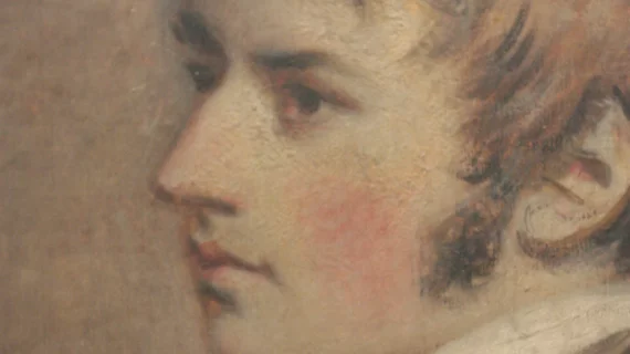
Scientists at the University of Bradford in England utilized medical imaging to analyze a painting believed to be by 19th-century artist John Constable.
Conducted on a privately owned piece to confirm its authenticity, the 19th-century oil painting underwent a series of non-destructive tests, including a CT scan, Raman spectroscopy, 3D microscopy and an X-ray, all to identify its chemical composition and any layers of paint.
The painting exhibited pigments typical of Constable's palette, and there seemed to be evidence of multiple layers, which is consistent with Constable's characteristic technique of "overwork," wherein the painting is re-worked at different times. However, the investigative process—which took several weeks—will take months of data analysis to confirm any findings. The results will then be presented to Constable experts for review.
Determining the actual painter behind a piece cannot be definitely confirmed through any analysis, but with enough evidence showing consistency with an artist’s other works, experts can make an educated determination. Regardless, the combination of techniques used for this analysis is largely unprecedented and could mark the most precise scientific study of a painting to date, according to coverage in the Yorkshire Post.
John Constable was renowned for his landscapes of the English countryside. He passed away in 1837, and his work is extremely valuable to collectors and historians alike.
Read the full feature from the Yorkshire Post at the link below.

