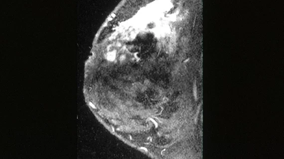
For high-risk women, even small lesions on breast MRI call for prompt assessment and biopsy
A U.K. study based on a departmental audit has confirmed previous research suggesting that MRI-detected small enhancing masses and new small enhancing foci, including those smaller than 5 millimeters, should be considered suspicious in women at high risk for breast cancer.
The new research was published online Dec. 5 in Clinical Radiology.
Anthony Maxwell, MB, ChB, of the University Hospital of South Manchester (and vice chair of the British Society of Breast Radiology) and colleagues queried the institution’s family-history database for women who had undergone screening MRI and been diagnosed with breast cancer within two years of the MRI exam.
The team found that, of 23 women who both met the criteria and had MRI images available for review, 14 were diagnosed at MRI, four at interim mammography, two symptomatically, one incidentally on ultrasound and two at risk-reducing mastectomy.
Ten of the 23 women (43 percent) had potentially avoidable delays in diagnosis.
The preceding MRIs were classified as false-negative screens in five women (one prevalent, four incident), false-negative assessment in seven and minimal signs in three (three women were assigned dual classifications), the authors report.
Common reasons for diagnostic delay included small enhancing masses that were overlooked, areas of non-mass enhancement that showed little or no change between screens, false reassurance from normal conventional imaging at assessment, and over reliance on short-interval repeat MRI.
Drawing from these findings, Maxwell and team offer several observations and recommendations on MRI breast screening in high-risk women, including:
- Small enhancing foci, masses, and areas of segmental non-mass enhancement are common MRI features of early breast cancer.
- Lack of change of non-mass enhancement on serial examinations does not exclude malignancy.
- Double reading of both screening and assessment examinations is recommended.
- Ready access to MRI biopsy is essential.
- Short-interval repeat MRI should be limited to reassessing low suspicion areas likely to be benign glandular enhancement.
- Annual mammography remains important in these women.
In their discussion, the authors state that their study emphasizes the importance of attention to detail in the technical performance and interpretation of screening breast MRI exams in high-risk women.
“Early rescanning of probably benign areas of non-mass enhancement may be appropriate, but new, persisting, or suspicious non-mass enhancement and new and indeterminate/suspicious masses or enhancing foci (even if small) should undergo prompt further assessment and biopsy,” they write.
Maxwell et al. further urge easy access to MRI-guided biopsy and recommend double-reading of screening and assessment imaging.
“Annual mammography appears to retain an important role, at least in older women, although its optimal timing in relation to MRI is uncertain,” they conclude.

