
MRI technique makes breast cancer 'glow' on imaging
A new MRI technique is said to be able to more accurately detect breast cancer by “lighting up” malignant tissue on images.
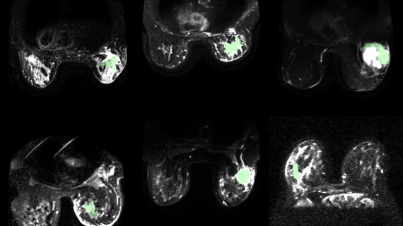
Developed by experts at the University of Waterloo in Ontario, Canada, the synthetic correlated diffusion imaging (CDI) technique highlights differences in how water molecules move through cancerous tissue in comparison to healthy tissue. This is achieved by synthesizing and mixing MRI signals at different gradient pulse strengths and timings.
Experts believe the technique can do more than just enhance cancer detection but improve disease management and treatment as well.
“This technology has great potential to not only improve breast cancer detection, but also treatment,” Alexander Wong, PhD, a professor in the Department of Systems Design Engineering at the University of Waterloo and co-director of the Vision and Image Processing (VIP) Lab, said in a release. “Our images contain key predictive information to help clinicians determine the best courses of action for treating each patient.”
The artificial intelligence-enabled system was first used for fine-tuning the detection of prostate cancer on MR imaging. To adapt the technique to breast cancer, researchers trained it using a diverse set of pre-treatment images from 350 breast cancer patients across 10 medical institutions with help from the American College of Radiology Imaging Network.
Aside from enhancing cancer detection, it is the team’s hope that by lighting up cancerous tissue, surgeons will be able to better visualize tumor margins. This could enable them to remove all cancerous tissue in a single surgery, rather than multiple, which is often the case. Clinical studies thus far have shown the CDI system to be incredibly effective for delineating tumor margins, routinely outperforming other gold-standard modalities in doing so.
“The more accurate we make CDI for delineating between cancerous tissue and healthy tissue, the more effective patient treatment plans and treatment itself can be,” Amy Tai, a Waterloo engineering PhD student supervised by Wong at the VIP Lab, said in the same release.
Given the success the team has observed in deploying the system in both breast and prostate cancer settings, the team is hopeful that they can continue to expand its utility to other types of cancer.
Learn more about the technique here.

In addition to her background in journalism, Hannah also has patient-facing experience in clinical settings, having spent more than 12 years working as a registered rad tech. She began covering the medical imaging industry for Innovate Healthcare in 2021.