
New low-field scanner detects cancer spread better than traditional breast MRI
A new type of MRI scanner may offer radiologists even better insight into the extent of patients’ cancer.
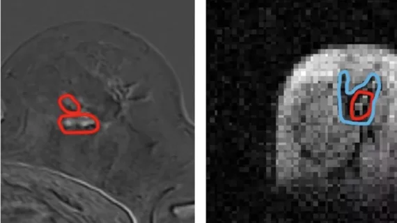
Developed by experts at the University of Aberdeen in Scotland in collaboration with National Health Service (NHS) Grampian, the Field Cycling Imager (FCI) scanner provides greater visualization of breast tissue in comparison to traditional breast MRIs. Researchers involved in its development are hopeful that the scanner could eventually lead to improved outcomes in cancer patients who require surgery to remove malignant tissue.
The scanner is capable of operating at different magnetic field strengths, including at ultra-low fields as low as 0.2T. This enables it to use multiple magnetic fields during a single exam, measuring the longitudinal relaxation time across a range of magnetic field strengths and giving providers additional information about the molecular properties of tissues.
Research into the use of FCI scanners revealed the technology to be particularly beneficial for analyzing breast cancer, as it can clearly differentiate tumors from adipose and glandular tissues without contrast agents. It can also ably distinguish between invasive and noninvasive cancers.
“We found that images generated from FCI can characterize breast tumors more accurately,” Lionel Broche, PhD, senior research fellow in Biomedical Physics and lead researcher in the study, said in a release on the team’s work. “This means it could improve the treatment plan for the patients by improving the accuracy of biopsy procedures by better detecting the type and location of tumors, and by reducing repeated surgery. So really, the potential impact of this on patients is extraordinary.”
Broche and colleagues believe their scanner could help reduce the likelihood that women who undergo lumpectomy will require multiple surgeries. Up to 15% of women who have lumpectomy require a second surgery to fully resect malignant tumor margins that are not completely removed during their first surgery.
“This is a truly exciting innovation and as we keep improving the technology for FCI, the potential for clinical applications is limitless,” Broche said.
While the findings thus far are promising, the team acknowledges that they need more prospective evidence to validate their data.
Learn more about the technology here.

In addition to her background in journalism, Hannah also has patient-facing experience in clinical settings, having spent more than 12 years working as a registered rad tech. She began covering the medical imaging industry for Innovate Healthcare in 2021.