Medical Imaging

Physicians utilize medical imaging to see inside the body to diagnose and treat patients. This includes computed tomography (CT), magnetic resonance imaging (MRI), X-ray, ultrasound, fluoroscopy, angiography,  and the nuclear imaging modalities of PET and SPECT. 

A study published this week in the Journal of the American College of Cardiology (JACC): Cardiovascular Imaging shows artificial intelligence (AI) algorithms can more rapidly and objectively determine calcium scores in computed tomographic (CT) and positron emission tomographic (PET) images than physicians.[1] The AI also performed well when the images were obtained from very-low-radiation CT attenuation scans. https://doi.org/10.1016/j.jcmg.2022.06.006

Artificial intelligence can objectively determine cardiac calcium scores faster than doctors

A new study shows artificial intelligence (AI) algorithms can more rapidly and objectively determine calcium scores in CT and PET/CT images than physicians.

Thumbnail

Deep learning reconstruction levels playing field between 1.5T and 3T MRI exams

Denoising using deep learning techniques can boost the performance of 1.5T MR brain imaging, resulting in quality comparable or superior to 3T imaging. 

Thumbnail

'Radiologic serendipity' a common trigger event that ends in thyroid surgery in asymptomatic patients

In this cohort, experts found that just 34% of surgeries were performed on symptomatic patients, which shifted their focus to other potential modes of detection.

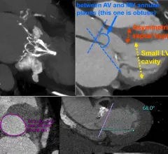
Examples of structural heart transcatheter valve replacement procedure planning CT scans and post procedure followup for TMVR and TAVR.

VIDEO: CT imaging for TAVR and TMVR structural heart interventions

Joao Cavalcante, MD, director, cardiac MRI and structural CT labs, Minneapolis Heart Institute, discusses the use of cardiac CT imaging to plan and guide structural heart procedures. 

coronavirus COVID-19 vaccine vaccination

MRI analysis offers new insight into vaccine-related lymphadenopathy in the general population

To date, research pertaining to reactive lymphadenopathy has focused mostly on patients with cancer who are routinely staged and monitored via PET/CT or in women undergoing breast cancer screening and/or imaging with mammography and ultrasound.

contrast enhanced mammography

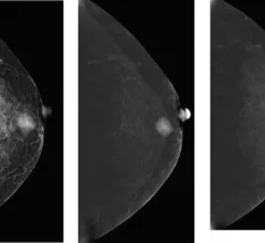
Scoring system outperforms BI-RADS assessments of contrast-enhanced mammography exams

For specificity—a metric that has been somewhat unpredictable in breast MRI and CEM—the scoring system performed significantly better than BI-RADS.

Thumbnail

Does the use of PSMA PET impact tumor upstaging?

A new risk analysis suggests the exam will increasingly result in tumor upstaging among high-risk patients.

Thumbnail

AI identifies pancreatic cancer frequently missed on CT

Specifically, the computer-aided detection (CAD) tool is capable of identifying lesions that are less than 2 cm.

Around the web

GE HealthCare designed the new-look Revolution Vibe CT scanner to help hospitals and health systems embrace CCTA and improve overall efficiency.

Clinicians have been using HeartSee to diagnose and treat coronary artery disease since the technology first debuted back in 2018. These latest updates, set to roll out to existing users, are designed to improve diagnostic performance and user access.

The cardiac technologies clinicians use for CVD evaluations have changed significantly in recent years, according to a new analysis of CMS data. While some modalities are on the rise, others are being utilized much less than ever before.