Society of Cardiovascular Computed Tomography (SCCT)

Around the web

Many new imaging solutions were on display at the world's largest health informatics conference, held at McCormick Place in Chicago.

Radiologist David Gruen, MD, discusses some of the overarching themes from the Healthcare Information Management Systems Society's annual meeting.

End-users of various radiology IT systems offer their assessment of the software in the annual KLAS Research 2023 Best in KLAS report.

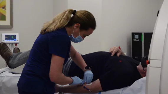
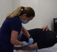
A CT coronary calcium scoring exam at Northwestern Medicine's Central DuPage Hospital in the Chicago Suburbs. Dave Fornell

The remaining gaps in evidence for cardiac CT

While the ACC/AHA 2021 Chest Pain Assessment Guidelines included cardiac CT angiography as a top level recommendation, gaps in evidence still need to be filled.

February 10, 2023
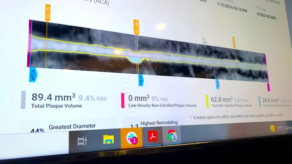
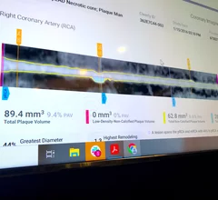
An example of an FDA cleared radiology AI algorithm to automatically take a cardiac CT scan and identify, contour and quantify soft plaque in the coronary arteries. The Cleerly software then generates an automated report with images, measurements and a risk assessment for the patient. This type of quantification is too time consuming and complex for human readers to bother with, but AI assisted reports like this may become a new normal over the next decade. Example from Cleerly Imaging at SCCT 2022.

Legal considerations for artificial intelligence in radiology and cardiology

There are now more than 520 FDA-cleared AI algorithms and the majority are for radiology and cardiology, raising the question of who is liable if the AI gets something wrong.

February 3, 2023

Increased use of CCTA improves CAD outcomes without raising costs

Researchers examined data from nearly 2 million patients, sharing their full findings in JACC: Cardiovascular Imaging.

January 23, 2023
Ed Nicol, MD, MBA, FSCCT, consultant cardiologist, honorary senior clinical lecturer, Kings College London, and president-elect of the Society of Cardiovascular Computed Tomography (SCCT), discusses what he sees as the big technology advances in cardiac CT. #SCCT #SCCT22 #RSNA22

VIDEO: New cardiac CT advances to watch

Ed Nicol, MD, MBA, president-elect of the Society of Cardiovascular Computed Tomography, discusses what he sees as the big technology advances in cardiac CT.

October 28, 2022
Ricardo Cury, MD, MBA, MSCCT, chairman of radiology, direct of cardiac imaging, Baptist Health South Florida and Miami Cardiac and Vascular Institute, discusses the new CAD-RADS 2.0 cardiac imaging reporting criteria at the 2022 SCCT meeting. Interview with Radiology Business Editor Dave Fornell.

VIDEO: What is new with CAD-RADS 2.0 cardiac imaging reporting?

Ricardo Cury, MD, chairman of radiology and director of cardiac imaging, Baptist Health South Florida and Miami Cardiac and Vascular Institute, discusses the new CAD-RADS 2.0 cardiac imaging reporting criteria.

October 26, 2022
AI-generated coronary tree from a patient's CT scan showing a color code of areas of interest for plaque burden from the Cleerly software shown at SCCT 2022.

VIDEO: The role of AI in cardiac imaging

Ed Nicol, MD, president-elect of the Society of Cardiovascular Computed Tomography, provided us with an exclusive look at how AI is expected to change cardiac imaging.

October 24, 2022
Coronary CT Angiography (CCTA) has evolved into a much more helpful resource than interventional cardiologists may have originally believed. In fact, the Society of Cardiovascular Computed Tomography (SCCT) emphasized in a new expert consensus statement that CCTA provides specialists with an effective tool in the preparation and optimization of coronary procedures. The 2021 chest pain guidelines elevated CTA to a class 1A recommendation. Harold Litt , MD, explains the use of CCTA in the ED.

VIDEO: Cardiac CT as a front-line chest pain exam in the ED

Harold Litt, MD, explains the use of cardiac CT as a primary imaging exam for chest pain at his emergency department over the past decade.

October 13, 2022
CT has been used for planning and procedural guidance in pulmonary vein isolation (PVI) for about 20 years. It shows the anatomy to the electrophysiologist. The EPs also can load the imaging into their EP mapping system. It also shows them the location of the esophagus and the phrenic nerve so they can avoid these during the procedures. #PVI #EPeeps

VIDEO: The expanding role of cardiac CT in electrophysiology

Harold Litt, MD, division chief of cardiothoracic imaging and director of the Center for Advanced CT Imaging Sciences at the Perelman School of Medicine of the University of Pennsylvania, explains the role of cardiac CT in EP procedures.

October 12, 2022
Trimed Popup
Trimed Popup