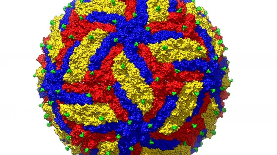
High-resolution image may be key to treating Zika virus
Researchers have captured the highest-resolution image to date of the Zika virus, which caused a global health crisis in 2015 leaving thousands of babies with serious birth defects. The research team believes the finding may aid in designing a vaccine to fight the virus.
The team used electron microscopy to create a 3D image of Zika, allowing them to visualize the atomic details of the virus. Results were published online June 26 in the journal Structure.
Compared to related viruses such as dengue and yellow fever, Zika is inherently more stable. This new 3D image allowed scientists to identify potential drug-binding pockets on the surface of the virus, which could aid the understanding of how vaccines interact with the virus.
“With the higher resolution, it is now possible to efficiently design vaccines and engineer anti-viral compounds that inhibit the virus," said corresponding author, Michael G. Rossman, a structural biologist at Purdue University in a release.

