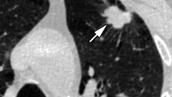
AI reduces CT lung cancer screening workload by nearly 80%
Artificial intelligence can significantly reduce the lung cancer screening-related workloads of radiologists.
CT screenings are integral to the early detection of lung cancer. However, identifying and segmenting lung nodules on imaging can be time consuming, adding to the reading burden for physicians. Many experts believe the emergence of AI applications could help address the issue.
New data published in the European Journal of Cancer details the potential AI has to completely change the workflows of providers who read lung cancer screenings. And it can do so with 100% accuracy as a first reader, according to the analysis.
The study was led by researchers at the University of Liverpool and the Research Institute for Diagnostic Accuracy in the Netherlands. Together, the team tested a tool developed by Coreline Soft, South Korea, on a dataset of over 1,200 CT lung cancer screenings. The group compared the AI’s performance to that of radiologists, assessing both readers’ accuracy based on positive and negative predictive values. Interpretation times also were evaluated to calculate time savings.
Compared to the reference standard, AI yielded fewer misclassifications of nodules compared to human readers, achieving a negative predictive value of 99.8%. All cancers that were histologically confirmed had been flagged by AI for further review, meaning that it didn’t miss any diagnoses.
Perhaps more impressive is the AI tool’s impact on workloads—the team estimated that the AI could reduce reading burdens by nearly 80%.
“Implementing low-dose CT screening for lung cancer is highly beneficial, but it comes with logistical and financial challenges,” John Field, PhD, lead author and Professor of Molecular Oncology at the University of Liverpool, and colleagues explained. “Our research suggests that AI could play a crucial role in making screening programs more efficient while maintaining diagnostic confidence.”
The group suggested that their findings indicate the algorithm has “considerable potential” to be implemented as a first reader.
Learn more about the team’s work here.

