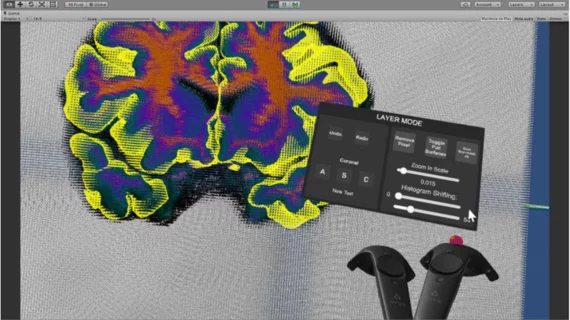
VR software may bring MRI segmentation into the future
A new virtual-reality (VR) software to correct segmentation errors on MRI scans was found to be faster, more accurate and enjoyable compared to a more commonly used system, report authors of a recent Journal of Digital Imaging study.
Manual segmentation is the current gold standard, according to lead author Dominique Duncan, with Keck School of Medicine at the University of Southern California in Los Angeles, and colleagues, but the process is time-consuming. The field has turned to automated segmentation as a solution.
“Correcting segmentation errors has been an important area of interest in neuroimaging for several decades, but despite significant research efforts, automated segmentation protocols are still imprecise and inaccurate,” Duncan and colleagues wrote. “Developing efficient segmentation correction methods could aid volumetric analysis of structures implicated in neurological disorders.”
Researchers created their own software—Virtual Brain Segmenter (VBS)—which utilizes a head-mounted VR system and a user-friendly platform.
Duncan and colleagues had 30 participants correct a single error on an MRI scan that had been previously segmented. They used either an open-source software using a traditional 2D interface or the Virtual Brain Segmenter. Prior to the process, each individual watched a five-minute demonstration on how to pick out and remove voxels representing the optic nerve on MRI images, and received five minutes of practice identifying the nerve.
Results showed those using the VR software completed segmentation 68 seconds faster than the 2D software (163.8 seconds compared to 231.8, respectively).
The VR method also achieved 85 percent precision and 86 percent sensitivity, each higher than a previous study using the 2D interface done by Ganz et al.
Twenty-eight participants completed a follow-up survey. All stated they preferred using VBS to the conventional 2D platform. Authors noted many said the VR system made segmenting “more exciting and fun.”
"We see this tool as potentially quite useful for correcting automated segmentation errors more effectively than a typical 2D screen/cursor interface," authors wrote. "We hope this study will be the beginning of a multistage project to create a gamified virtual reality program that can be utilized by a crowd to correct segmentation errors."

