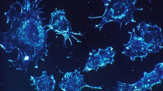
Molecular imaging tool can map cancer cell division in real-time
Researchers from Columbia University in New York have developed a molecular imaging tool that can track metabolic changes in individual living cells in real time.
The research, published July 30 in Nature Communications, details how the tool combines the chemical tracer D20, or heavy water, with a new laser-imaging method called stimulated Raman scattering (SRS) to label lipids and proteins to track subcellular changes.
For their study, the researchers diluted regular water with D20 and administered it to roundworms, mice and zebrafish embryos to drink. They then aimed the SRS laser at different tissue and examined deuterium-tagged proteins, lipids and DNA build up inside the animals' cells.
In some instances, the researchers were able to see a bright line emerge around dividing cancerous cells in tumors present in the brain and colon and more deuterium was incorporated into their newly made proteins and lipids, according to the researchers.
The technique may help surgeons remove tumors quicker and detect tumors, head injuries or developmental and metabolic disorders sooner, explained senior author Wei Min, PhD, a chemistry professor at Columbia.
"We can use this technology to visualize metabolic activities in a wide range of animals," Min said in a prepared statement. "By tracking where and when new proteins, lipids and DNA molecules are made, we can learn more about how animals develop and age, and what goes wrong in the case of injury and disease."

