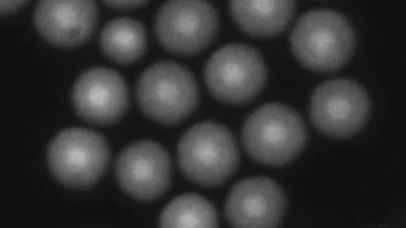
Light-emitting nanoparticles may provide safer deep-tissue imaging, guide radiation therapy
Researchers from the Department of Energy's Lawrence Berkeley National Laboratory have developed an imaging application that utilizes light-emitting nanoparticles and could provide a safer way to see deeper into living tissue and cells, according to an Aug. 15 news release.
The study, published online Aug. 6 in Nature Communications, details how the researchers designed alloyed upconverting nanoparticles (UCNPs) to activate from ultralow power laser light at near-infrared wavelengths. The UNCPs can absorb this light and then emit visible light that can be measured by standard imaging equipment.
Some imaging systems use high-power laser light that can damage living cells in the human body. Co-lead author Bruce Cohen, PhD, a staff scientist with the University of California, Berkeley, and colleagues, however, asserted that their imaging application is safer because the laser power needed for UCNPs is exceedingly lower than the power needed for conventional near-infrared-imaging probes.
For their study, Cohen and colleagues injected nanoparticles into the mammary fat pads of mice and recorded images of light emitted by the particles. The UCNPs were small enough to penetrate the tissue, measuring 12 to 15 nanometers across, according to the researchers.
The researchers observed that this process did not harm or toxify the mice cells, however they explained that additional research is needed to know if the technique is safe for humans and to test coatings being designed to specifically bind to cancer cells. Nonetheless, the system may help guide intensive surgeries and radiation treatment.
“This new paradigm in UCNP design, which leads to much brighter particles, is a real game-changer for all single-UCNP imaging applications,” said researcher Jim Schuck, PhD, a former Berkeley Lab scientist now at Columbia University, in a prepared statement.

