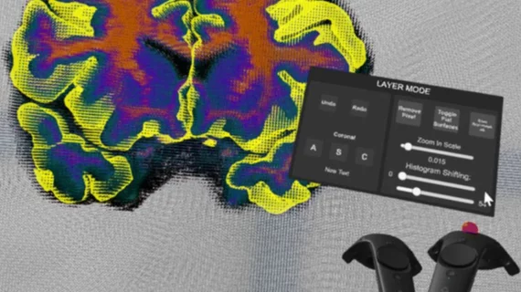
VR tool could enhance medical imaging segmentation, error correction
Medical imaging data may become an entirely immersive experience with the development of a new virtual reality (VR) tool created by researchers at the University of Southern California (USC) Keck School of Medicine.
Virtual Brain Segmenter (VBS) is a tool that automatically processes medical imaging data such as MRIs through segmentation. It could increase the efficacy of brain scan analysis, according to a USC news release from Aug. 21.
Researchers used VBS with MRI data to separate the brain into labeled regions to help with examination of various structures, as described in a study published online in the Journal of Digital Imaging.
Researchers—led by Dominique Duncan, MD, assistant professor of neurology at the Keck School of Medicine—were motivated to make the segmentation process more efficient by finding a way to correct errors that requires less training.
“Segmentation of MRI scans is a critical part of the workflow process before we can further analyze neuroimaging data,” the researchers wrote. "Although there are several automatic tools for segmentation, no segmentation software is perfectly accurate, and manual correction by visually inspecting the segmentation errors is required."
For the project, Duncan and colleagues teamed with RareFaction Interactive, which programmed and designed VBS, and secured a Vive VR headset from electronics company HTC.
Thirty participants who had never performed brain segmentation were given the VR headset and two controllers. Each participant was tested using a MacBook to perform minor error correction in the FreeSurfer data cleanup program. They then completed the same task in VBS, according to the researchers.
The researchers found that participants using VBS finished the correction 68 seconds more quickly than those using FreeSurfer, who took more than three minutes to complete.

