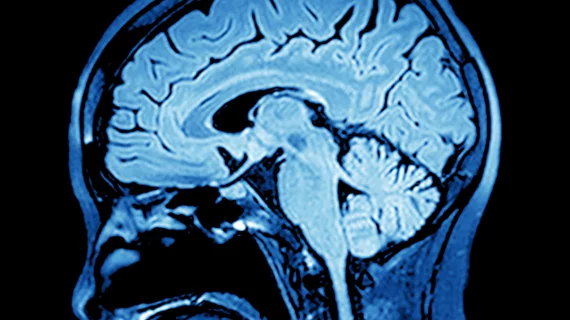
Brain scan distinguishes between bipolar disorder, depression
Functional MRI (fMRI) may be the key to identifying specific neurons in the brain that are central to distinguishing bipolar disorder from depression, reported researchers in a recent Biological Psychiatry: Cognitive Neuroscience and Neuroimaging study.
"Mental illness, particularly bipolar disorder and depression, can be difficult to diagnose as many conditions have similar symptoms," said lead author Mayuresh S. Korgaonkar, MD, in a release. "The wrong diagnosis can be dangerous, leading to poor social and economic outcomes for the patient as they undergo treatment for a completely different disorder.”
Researchers, led by a team from the Westmead Institute for Medical Research at the University of Sydney in Australia, employed fMRI to visualize how the amygdala reacted as 81 patients processed anger, fear, sadness, disgust and happiness. Thirty-one patients had remitted Bipolar I disorder, 25 with remitted major depressive disorder and a further 25 in a healthy control group.
Overall, bipolar participants had a less active and less connected left amygdala compared to the 25 unipolar major depressive disorder patients. The method was 80 percent accurate in distinguishing between the two.
“These findings suggest that differences in amygdala activation and connectivity during processing of facial emotions persist beyond the depressed and manic states and potentially could be a trait marker for differentiating these disorders,” Korgaonkar et al. wrote in the study.
The team’s testing did not involve patients who were either currently experiencing manic or hypomanic symptoms nor currently depressed, they noted as a limitation of the study. However, some measures correlated with the total number of episodes and illness age of diagnosis, the team wrote, lending support for “trait-like characteristics.”
Korgaonkar and colleagues are currently conducting phase two of the study, according to the release, in an attempt to further characterize the markers of bipolar disorder and depression in a larger group of patients.

