Electron microscopic imaging of virus may reveal new potential for gene therapy
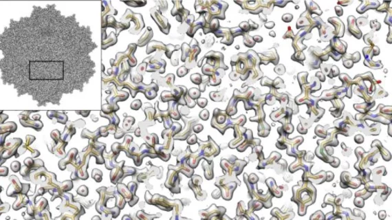
Researchers from the Salk Institute in San Diego, California and the University of Florida have used cryo-electron microscopy (cryo-EM) imaging to analyze a three-dimensional model of the molecular structure of the AAV2 virus. The advanced molecular imaging technique may demonstrate the virus’ potential as a delivery vehicle for gene therapies, according to research published Sept. 7 in Nature Communications.
"We applied a number of different procedures that have previously only been described in theory,” said co-author Dmitry Lyumkis, PhD, an assistant professor at Salk, in a prepared statement. “We demonstrated experimentally, for the first time, that they can be used to dramatically improve the quality of this kind of imaging."
Lyumkis and colleagues focused their research on a single strain of an AAV2 virus that has a distinct change in one of its amino acids, causing it to be less infectious than others.
The researchers were then able better understand the interactions between different viruses and the types of cells they infect to see how the immune system recognizes viruses.
The high level of symmetry of the virus also made applying computation techniques ideal for the cryo-EM analysis, according to Lyumkis.
"We were able to get 60-fold more information, which allows us to apply new computational techniques to create a better reconstruction of the molecule that will be extendible to many future high-resolution cryo-EM experiments,” Lyumkis said.
The results of the study show the potential to use lower-voltage microscopes for data generation techniques and enable researchers to use more cost-effective imaging tools in the future.

