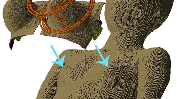
Simulation reveals how breast tissue of varying density may react to MRI
Novel computer simulations of the female body developed by researchers from Purdue University in West Lafayette, Indiana, may help predict how more than 20 different breast tissue ratios will respond to MRI varying in radiofrequency.
Currently, there is no way to clinically prove that an MRI technique is safe for all women of varying breast densities before starting a clinical trial.
However, the newly developed computer simulation model may show whether MRIs meet FDA safety guidelines, according to research published online Sept. 14 in Magnetic Resonance in Medicine.
“We're starting to develop techniques at high field strengths that could immediately monitor how tumors respond to treatment. So, we don't want tissue heating concerns to stand in the way of improving such a powerful tool," said Joseph Rispoli, PhD, assistant professor of biomedical engineering at Purdue, in a prepared statement.
Rispoli and colleagues demonstrated through their virtual simulations that a five-fold increase in the specific absorption rate (SAR) could stay within FDA limits.
Due to the lack of full body female computer simulation models—phantoms—available, the researchers created their computer models by fusing 36 female phantoms with various breast densities as classified by the American College of Radiology Breast Imaging Reporting and Data System Atlas along with female phantoms created by researchers in Japan and Sweden.
They then simulated the behavior of each fused phantom in response to MRI coils at 7 tesla, which helped the researchers customize their techniques to each of the 20 different breast tissue ratios.
"We want to facilitate the most cutting-edge of breast MRI techniques at any site in the world," Rispoli said. "Ultimately, a woman will be able to go in, have a fast low-power anatomical MRI scan, and then the computer could quickly simulate on-the-fly what the SAR would be in that patient."

