Minimally invasive CT, MRI autopsy may enhance postmortem diagnoses
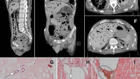
Minimally invasive autopsy using CT and MRI performs as well as conventional autopsy— though better determined unexpected causes of death and diagnostic information, according to a study published online Sept. 25 in Radiology.
"With minimally invasive autopsy, the body is investigated from top to toe, and with regular autopsy usually only the torso and, if consented by next-of-kin, the brain," said J. Wolter Oosterhuis, MD, PhD, from the Department of Pathology at Erasmus University Medical Center in Rotterdam, the Netherlands, in a prepared statement. "Moreover, postmortem CT and MRI demonstrate pathology, in particular of the skeleton and soft tissues, that is easily missed by the regular autopsy."
Oosterhuis and colleagues compared the performance of minimally invasive autopsy combining postmortem MRI, CT and CT-guided biopsies to conventional autopsy in 99 human corpses.
Overall, the methods similarly determined the immediate cause of death in 92 percent of cases, according to the researchers.
Additionally, there was more agreement with the cause of death in the minimally invasive autopsy case than with conventional autopsy. The total number of diagnoses determined by minimally invasive autopsy were higher than conventional autopsy — minimally invasive autopsy diagnosed 90 percent of diagnoses compared with 78 percent for conventional autopsy.
The researchers noted that minimally invasive autopsy also provides information that can be stored for long-term use unlike conventional autopsy.
"It's very important to note that conventional autopsy cannot be redone, and items that were overlooked or misinterpreted cannot be corrected," Oosterhuis said.
"Minimally invasive autopsy, on the other hand, provides a permanent record of the entire body that can be revisited, and reanalyzed by pathologists, radiologists, clinicians and next-of-kin," Oosterhuis added. "For scientists, this dependable database has great potential for future research."

