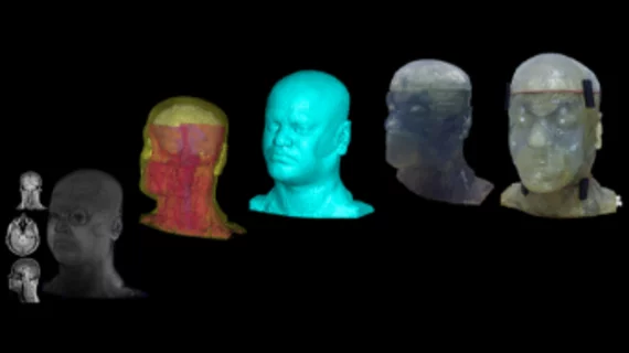
3D printed phantom head may improve MRI safety before human testing
A bioengineering doctoral student at the University of Pittsburgh has developed a 3D phantom human head to test MRI systems and other imaging technologies before testing on human subjects, according to a recent University of Pittsburgh press release.
Sossena Wood, and her advisor Tamer Ibrahim, PhD, an associate professor of bioengineering at the University of Pittsburgh, developed the phantom to better understand why moving from lower to higher magnetic resonance fields with 7T MRI produces images of varying quality and its affect on localized heating.
“We wanted to develop an anthropomorphic phantom head to help us better understand these issues by providing a safer way to test the imaging," Ibrahim said in a prepared statement. "We use the device to analyze, evaluate and calibrate the MRI systems and instrumentation before testing new protocols on human subjects."
The researchers built the digital blueprint for the 3D printed model using a 3T MRI dataset of a healthy male—characterizing it by segmentation and breaking it into eight tissue components to improve image accuracy. An MRI scanner was then used to produce the 3D digital image of the male’s head which was then printed as an anthropomorphic phantom head prototype.
The phantom can be used to see if implants are able to go inside of an MRI or detect the temperature rise in different bodily tissues based on various radiofrequency (RF) instrumentation, according to the release.
“With MR imaging, the power from the radiofrequency exposure is transformed into heat in the patient’s tissue, which can have detrimental effects on the patient’s health, especially with implants if not monitored by the scanner,” Wood said in a prepared statement. “With our phantom head, we can test the safety of our imaging by putting probes inside of certain regions of the head and measuring the effects.”

