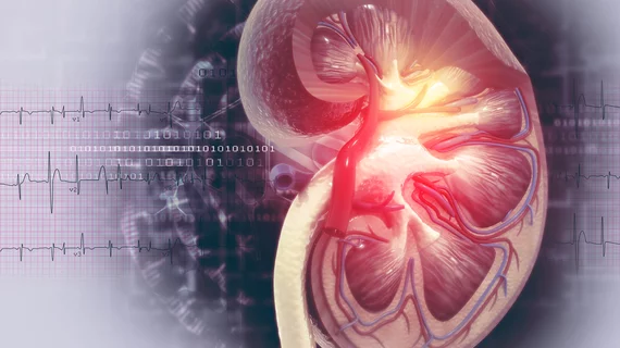
Microscopic imaging of kidney damage from contrast dyes opens door for novel drug therapies
Using microscopic imaging, researchers from the University of Calgary in Alberta, Canada have shown how the kidneys negatively respond to contrast dyes used during various medical tests and procedures, according to a university press release published Oct. 15.
The findings—published in the June issue of the Journal of Clinical Investigation—may help physicians find new methods to better protect the kidneys and prevent patients from going on dialysis.
“This study is important because it increases our understanding of how we might intervene to prevent acute renal injury or interrupt the progression of acute kidney injury to chronic kidney disease thereby reducing the burden of kidney disease among Canadians,” Norman Rosenblum, MD, scientific director of the Canadian Institutes of Health Research (CIHR) Institute of Nutrition, Metabolism and Diabetes, said in the statement.
For their study, University of Calgary professors and kidney specialists Matthew James, MD, PhD, and Dan Muruve, MD, used specialized high-powered microscopes to map out in real time the progression of a contrast dye though the kidneys of mice.
The researchers found that whether the kidney was hydrated determined if the kidney flushed out the dye or absorbed it and became inflamed or damaged. These findings then were translated into a smaller study in which the researchers used human urine after contrast dye exposure. The results pointed to the same markers as in the mice, according to the researchers.
“These results can help us add to the steps we currently emphasize to reduce the amount of contrast dye used and to hydrate the patient,” James said in a prepared statement. “Despite this, some patients with kidney disease currently avoid these medical tests because of the concern about possible injury to their kidneys. This research could help make these tests even safer for them.”
The study has also helped the researchers develop new drug therapies for patients who have weak hearts and cannot ingest extra fluids to get hydrated.
“Through this research we’ve discovered a drug that stops the kidney from absorbing the dye to prevent possible injury,” Muruve said in the statement. “We’ve tested a medication that is showing promising results. The next step is to translate these findings into clinical trials.”
The research was supported by Canadian Institutes of Health Research (CIHR) and The Kidney Foundation of Canada.

