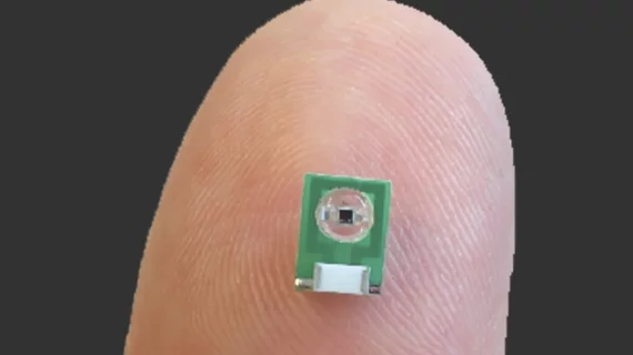
Micro MRI sensor can monitor electromagnetic brain activity
Engineers from MIT have developed a non-invasive MRI sensor no bigger than a penny that can detect and measure electrical activity or optical signals in the brain. The research was published online Oct. 22 in Nature Biomedical Engineering.
The sensor widens the capabilities of a standard brain MRI—which is commonly used to measure changes in blood flow that indirectly represent brain activity—by detecting electrical currents and light produced by luminescent proteins in the brain.
“MRI offers a way to sense things from the outside of the body in a minimally invasive fashion,” lead author Aviad Hai, a postdoctoral student at MIT, said in a prepared statement. “It does not require a wired connection into the brain. We can implant the sensor and just leave it there.”
To ensure their sensor could detect electromagnetic fields with accurate spatial resolution, the researchers inserted a tiny radio antenna directly into the brain to receive radio waves generated by hydrogen atoms in brain tissue.
Additionally, the sensor is self-sufficient and doesn't need to carry any kind of power supply because the radio signals emitted by the external MRI scanner are enough to power the sensor, according to the researchers.
The researchers then tuned their sensor to the same frequency as radio waves emitted by hydrogen atoms in the brain. When the sensor picked up an electromagnetic signal from the brain tissue, its tuning changed and the sensor no longer matched the frequency of the hydrogen atoms. In turn, this created a weaker image when the sensor was scanned by an MRI machine.
The sensor demonstrated that they could detect electrical signals like those produced by action potentials (the electrical impulses fired by single neurons), or local field potentials (the sum of electrical currents produced by a group of neurons), according to the authors.
To verify its accuracy, the sensors were used in a rat model to see if it could pick up signals in living brain tissue. The researchers found the sensor could precisely pinpoint cells engineered to express the protein luciferase that were emitting light.
In the future, the researchers hope to make their sensors even smaller. They also interested in using their sensor to detect neural signals in the brain and monitor other electromagnetic signals in the body, such as muscle contractions or cardiac activity.
“If the sensors were on the order of hundreds of microns, which is what the modeling suggests is in the future for this technology, then you could imagine taking a syringe and distributing a whole bunch of them and just leaving them there,” said senior author Alan Jasanoff, PhD, a professor of biological engineering, brain and cognitive sciences, and nuclear science and engineering, at MIT. “What this would do is provide many local readouts by having sensors distributed all over the tissue.”
The research was funded by the National Institutes of Health (NIH).

