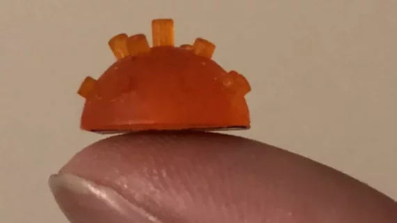
How a miniaturized pipe organ could improve ultrasound images
A miniaturized version of a musical instrument developed by researchers at the University of Strathclyde in Glasgow, Scotland, can’t produce actual music, but it may improve the quality of medical images, according to research published in the October issue of IEEE Transactions on Ultrasonics, Ferroelectrics, and Frequency Control.
The design of the miniaturized pipe organ is based on the actual wide range of pipes in the full-sized instrument. With this, the researchers designed the pipe organ, or ultrasonic transducer—used to receive and transmit ultrasonic signals—to improve images from scanners by broadening the range of frequencies used to emit sound waves.
“Musical instruments have a wide variety of designs but they all have one thing in common – they emit sound across a broad range of frequencies,” study author Tony Mulholland, PhD, chair of Strathclyde’s Department of Mathematics and Statistics, said in a prepared statement. “So, there is a treasure trove of design ideas for future medical imaging sensors lying waiting to be discovered amongst this vast array of designs.
Roughly 20 percent of medical scans are performed using ultrasound, Mulholland explained, and the scanner operating at a single frequency partly accounts for producing ultrasound images with poor resolution.
For the study, Mulholland and colleagues used a 3D printer to create the best designs for the transducer to produce multiple frequencies like an actual pipe organ. The designs were then tested using mathematical models and computer simulations.
“Developing wide bandwidth ultrasound systems could give significant improvements in imaging capability,” said co-author James Windmill, of Strathclyde’s Centre for Ultrasonic Engineering, PhD. “Using high resolution 3D printers allows us to try new, three-dimensional device designs with much faster development cycles.”
Though in its stages of early development the researchers hope the technology could aide in the design of hearing aids, underwater sonar and the non-destructive testing of nuclear plants, according to the release.

