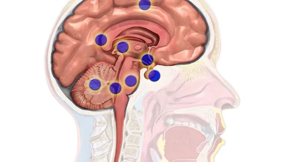
International team uses AI to uncover cellular makeup of various brain regions
With data obtained from fMRI scans and machine learning, National University of Singapore-led (NUS) researchers have a better understanding of the cellular architecture of the brain.
Performed in conjunction with researchers in the Netherlands and Spain, the team was able to automatically estimate measurements of the brain to calculate the cellular makeup of various brain regions. The non-invasive insights may be used to assess treatment of neurological disorders and to develop new therapies, according to authors of the Jan. 9 research published in Science Advances.
"The underlying pathways of many diseases occur at the cellular level, and many pharmaceuticals operate at the microscale level,” said Thomas Yeo, with the Singapore Institute for Neurotechnology at NUS, in a news release. “To know what really happens at the innermost levels of the human brain, it is crucial for us to develop methods that can delve into the depths of the brain non-invasively.”
Yeo and colleagues examined imaging data from 452 patients who were part of the Human Connectome Project. They discovered that parts of the brain involved in sensory perception—vision, hearing and touch—demonstrated characteristics considered opposite from regions involved in internal thought and memories.
According to the team, this cellular architecture pattern of the brain provides insight into how it hierarchically processes information.
"Our study suggests that the processing hierarchy of the brain is supported by micro-scale differentiation among its regions, which may provide further clues for breakthroughs in artificial intelligence," Yeo added.
The authors plan to apply their study to the brains of individual patients for a better understanding of cellular variation.

